South Africa and the Cradle of Sauropod-Kind

Artist's reconstruction of Pulanesaura by Gina Viglietti.

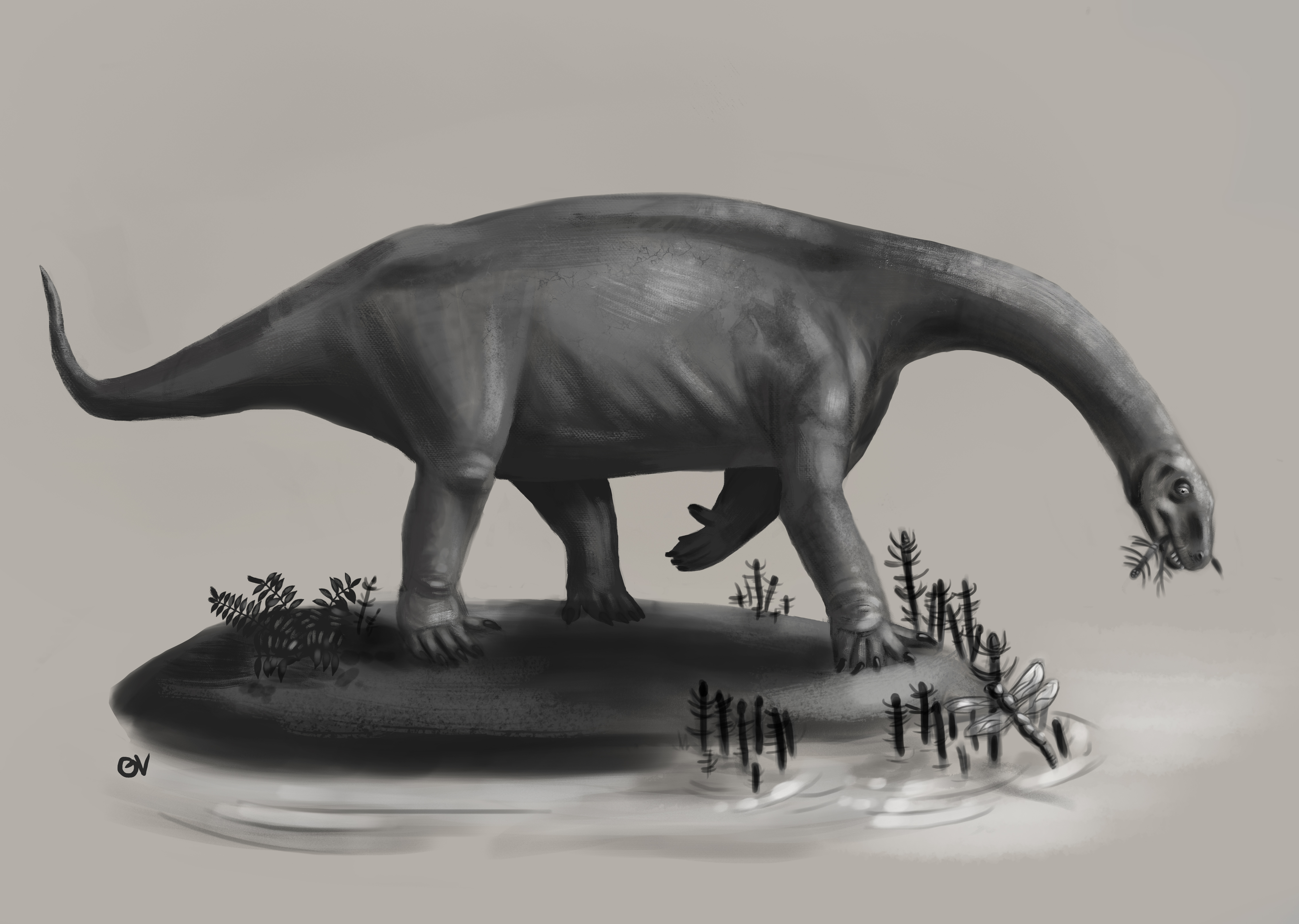
Artist’s reconstruction of Pulanesaura by Gina Viglietti.

I am happy to report on a new sauropod dinosaur from the Early Jurassic of South Africa! The dinosaur, Pulanesaura, was discovered in 2004 and has been a long time coming to press. Now, she’s finally here.

When I was maybe four or five years old, I remember reading a dinosaur book with my mother. The book described for children how dinosaurs were discovered and excavated in the field, and then how the bones were reassembled back in the lab. What I don’t remember, but I have been told, was that at some point during the explanation of how dinosaurs were unearthed, I interjected, “And then they have lunch.”

I was five years old in 1978. Fast-forward to the fall of 2004, and my 31-year-old self is standing in the rain at Spion Kop farm in the Free State of South Africa marveling at the large limb bones poking out of a section of the upper Elliot Formation. I don’t recall having lunch at that moment, but I do remember being excited, and being grateful that I was part of a team, headed by Adam Yates, charged with exploring these Early Jurassic rocks. National Geographic had sponsored our grant to pursue promising bones issuing forth from these rocks, and although we did not immediately know we had a new dinosaur, there was a good possibility we did.

Matt w tibia (1)

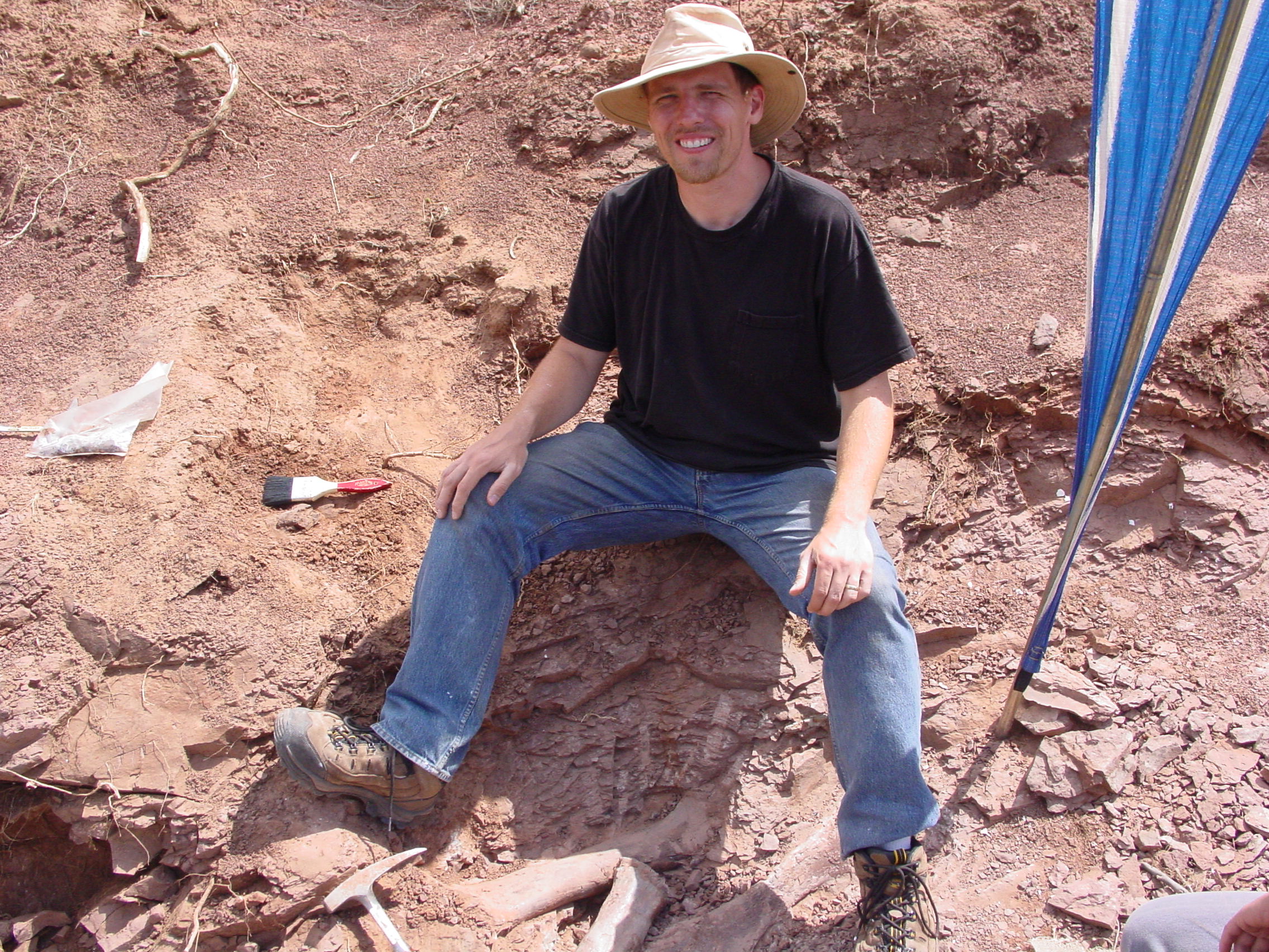
Myself above two tibia bones (tibiae) from what would become known as Pulanesaura in 2004 … it was a less rainy day that day.

Why South Africa? As it turns out, South Africa has many exposures of Lower Jurassic rocks that record a very significant time in dinosaur evolution. The earliest true dinosaurs appeared in the Triassic period about 235 million years ago (Ma) but remained relatively small to medium-sized animals that were in competition with other vertebrate groups vying for dominance in a harsh world. During the Triassic period, all the continents were amalgamated into a single supercontinent dubbed Pangea. Although to a modern traveler the thought of Pangea sounds amazing (imagine riding a train from North America over to Europe or driving from South America into Antarctica, Africa, or Australia), ecologically this was disastrous. A huge expanse of Pangea was landlocked and nearly devoid of water, making it both hot and uninhabitable. Moreover, sea levels were also drastically lower, creating fewer areas for marine life to thrive. Thus, Pangea took a huge toll on the animals that preceded the dinosaurs. In fact, the largest mass extinction in the past 540 million years occurred just prior to the Triassic period, wiping out a majority of life on the planet.

During the Early Jurassic (starting about 200 Ma), Pangea began to unzip and break up into separate landmasses, and part of the effect of this was to bring water into regions it hadn’t been in millions of years, supporting more plants and, in turn, the animals that fed on them. It is also at this critical juncture that the sauropodomorph dinosaurs began to become larger-bodied and more diverse so that by the end of the Jurassic period (about 145 Ma), many of these herbivores were tipping the scales at 20-30 metric tons! Sauropodomorphs started out as small to medium-sized bipedal herbivores that used their long necks and grasping hands to consume foliage at different heights in their environment. Sauropods became fully quadrupedal giants with elongate necks that acted as efficient food-gathering feeding booms, sweeping across swaths of vegetation while the herbivore stayed put. And this transition from mostly bipedal herbivores eating with their hands and necks to giant quadrupeds that relied solely on long necks to feed occurred right around the Early Jurassic period about 200 Ma. So, if you want to understand the beginnings of this trend towards gigantism in the sauropodomorph dinosaurs, you need to search for fossils in Early Jurassic rocks … and that brings us back to me standing in the rain at Spion Kop on the upper Elliot Formation staring at the large bones coming out of the ground.

Those bones we were unearthing would end up being a sauropod new to science named Pulanesaura that my colleagues and I have published on this week in Nature Scientific Reports. The lead author, Blair McPhee, is a Ph.D. student at the University of Witwatersrand in South Africa who took on this dinosaur for his dissertation. Remember that we discovered Pulanesaura in 2004?  Why didn’t we publish on this animal earlier? For a number of reasons collectively called life. Between 2009 and 2011, we did publish on two other sauropodomorph dinosaurs from Spion Kop, so that took up some time. But more significantly, Adam Yates and I had major life-changing moves to new employers: Adam to the Museum of Central Australia in Alice Springs; me to Stockton University. And so poor Pulanesaura was languishing. Therefore, when Blair approached us about describing Pulanesaura for his Ph.D., we were enthusiastically supportive. I was especially pleased to see Blair at the helm of the description. I am beyond happy that a South African Ph.D. student is the lead author on the description of a native South African dinosaur. His persistence and perseverance on this project is why the world now knows about Pulanesaura.

Why Pulanesaura? Well, the name means “rain bringer” in Sesotho, which is fitting since we always seemed to get rained on during the excavation of this dinosaur. And the publication of “Rain Bringer” has finally brought home a trilogy of sauropodomorph dinosaurs and the complex story they tell of what was happening in the Early Jurassic at what is now the Spion Kop farm.

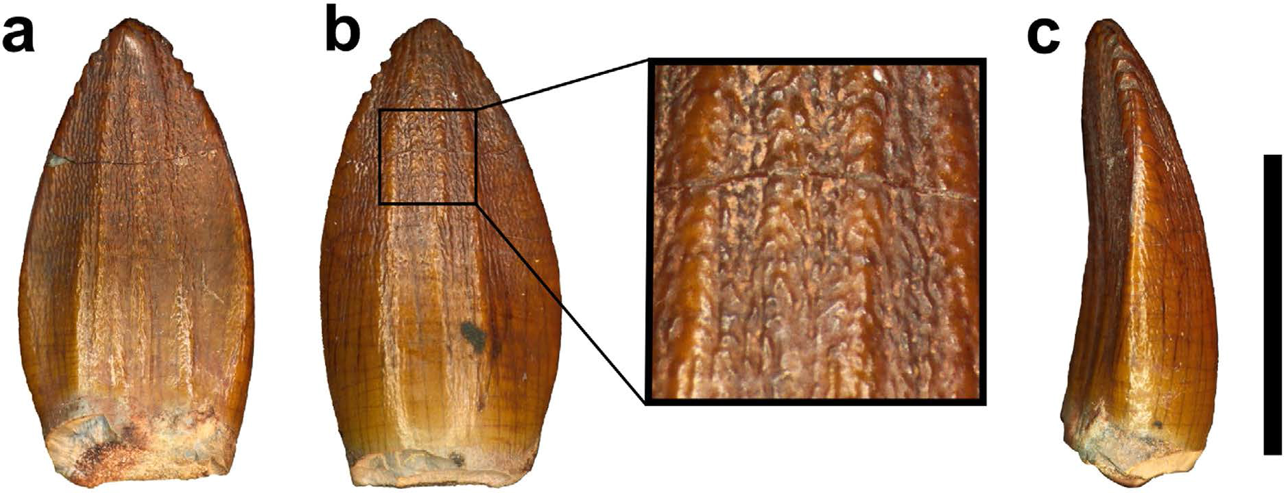
First things first – how do we know Pulanesaura is a sauropod? A number of clues point the way. For one thing, although we did not find a skull, we found teeth. The teeth of sauropods, unlike their sauropodomorph brethren, have a spoon- or spatula-like profile and have wrinkled enamel. The teeth of Pulanesaura certainly fit the bill there.

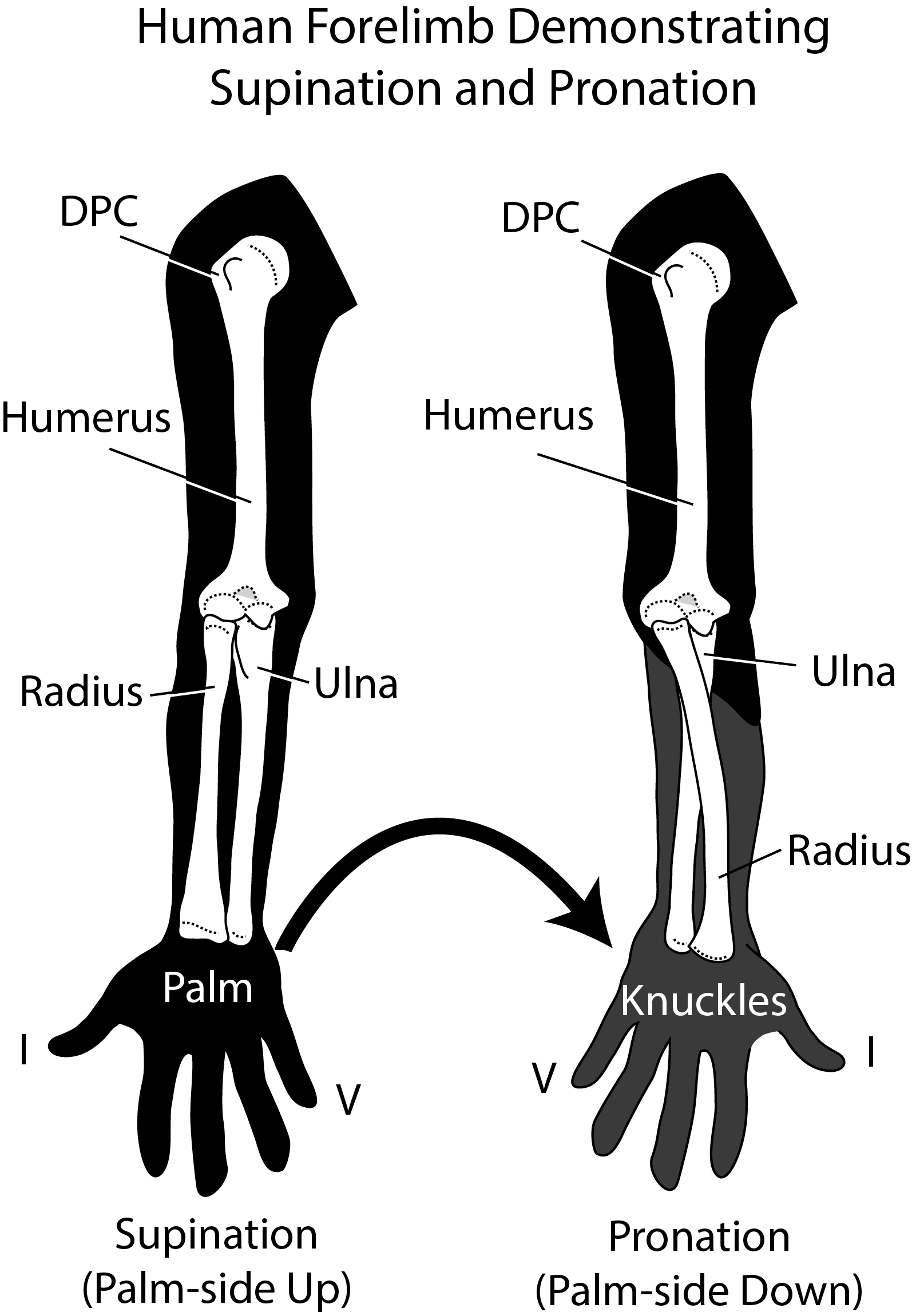
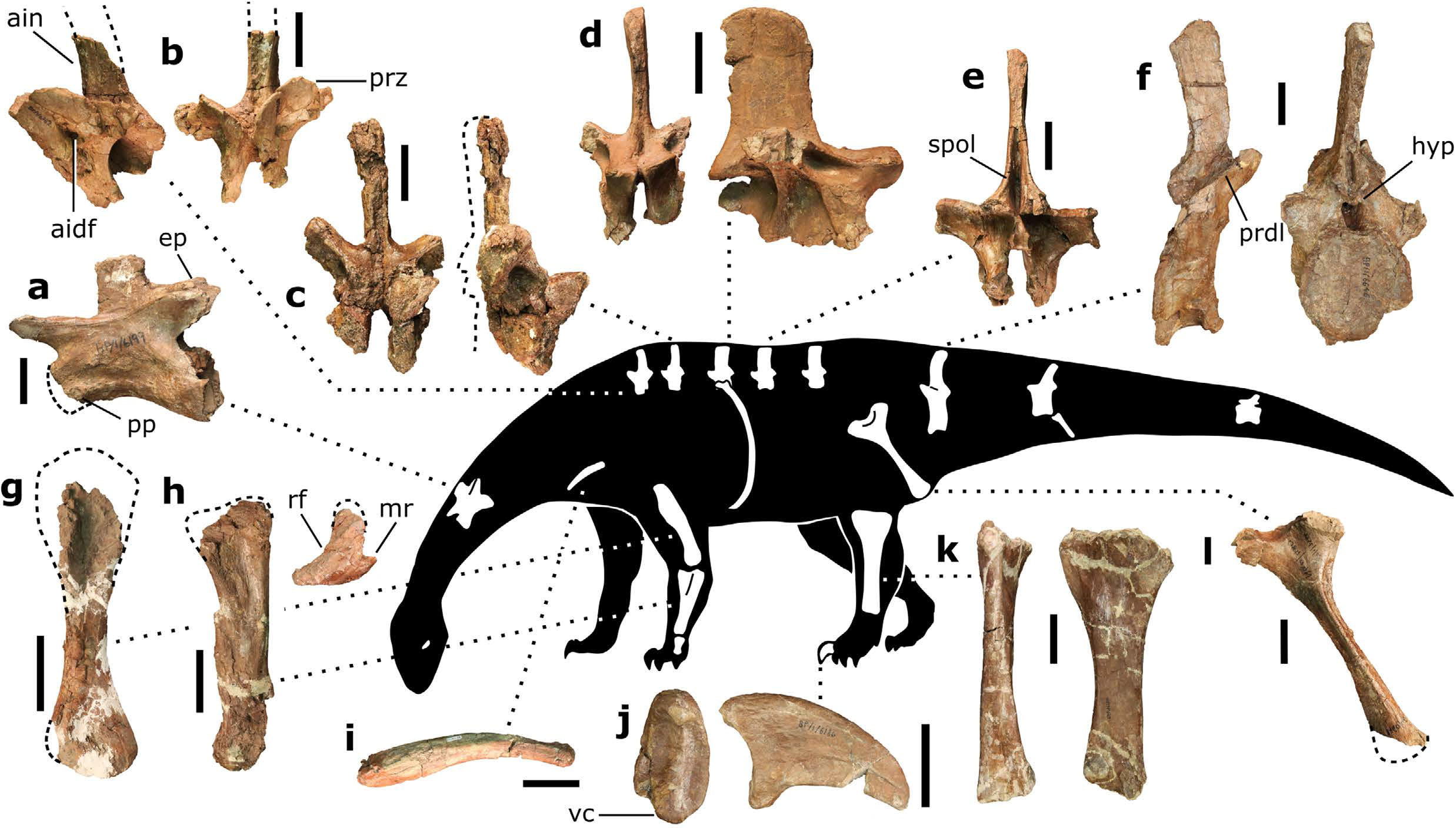
Next, being large, sauropods braced their vertebral column with extra joints in their backbones (vertebrae) – a portion of this extra joint is called the hyposphene. The body vertebrae we have of Pulanesaura only preserve their tops (the hyposphene is located on the bottom-half of the vertebra) but luckily a full tail (caudal) vertebra is preserved, and that has a hyposphene. We are also fortunate that part of the forelimb was preserved. The ulna bone in sauropods cradles the other forearm bone, the radius, by wrapping around it from behind. In sauropods, a wide, triangular depression is present on the ulna where the radius sits. Although the ulna of Pulanesaura is a bit crushed and scrappy, it was intact enough to show that, yes, indeed, such a depression for the radius was there. These features and more showed us that this dinosaur was certainly a true sauropod.

How do we know Pulanesaura is new to science? Using a method called cladistics, the suite of features for Pulanesaura was compared to other sauropodomorphs and sauropods from South Africa and around the globe. Its unique combination of features show that it is not a member of previously known sauropodomorph or sauropod dinosaurs, but falls along its own branch of the dinosaur family tree near the common ancestor of all sauropod dinosaurs. At the moment, it is very difficult to tell the difference between a true early sauropod and a sauropodomorph very close to the common ancestor of sauropods. Given the data we have for Pulanesaura, we find it most likely to be a very early sauropod. Certainly, future studies and perhaps more material of Pulanesaura will clarify this picture.

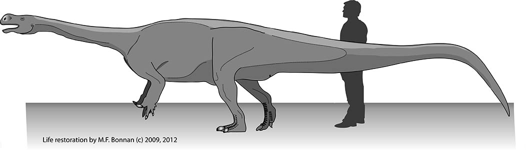
How big was Pulanesaura? We certainly don’t have a complete skeleton of this herbivore, but we have enough bones from enough areas of the body to infer that this animal stretched nearly 8 meters (about 26 feet) long and stood about 2 meters (about 6.5 feet) high at the hip. That may seem big, but it’s small for a sauropod.

Why is Pulanesaura significant? The traditional picture of sauropodomorph evolution is that when true sauropods came onto the scene, the other sauropodomorphs were pushed aside, their small body size and “inferior” anatomy undone by the larger herbivores. But Pulanesaurua turns this notion on its head because, living alongside it at Spion Kop were other sauropodomorph dinosaurs with very different anatomies. Adam, Johann, myself, and others have described two of these other sauropodomorphs, both also from Spion Kop. One, Aardonyx, was a 7 meter long sauropodomorph capable of assuming both a bipedal and quadrupedal posture; and another, Arcusaurus, was a small, juvenile sauropodomorph with a hold-over of more primitive features. And one of the things these other sauropodomorphs had going for them was that they could feed at different heights and use their forelimbs to direct foliage to the mouth. In contrast, the anatomy of Pulanesaura shows that it was an obligate quadruped (it could not stand bipedally on its hind legs), which would have restricted its vertical reach for vegetation compared with these other sauropodomorphs. However, the single neck (cervical) vertebra we have for Pulanesaura has joints that were spaced and angled (much like those of other sauropods) such that they would have allowed for a larger range of neck motion than in other contemporaneous sauropodomorphs. In other words, although Pulanesaura could not rear up and extend its neck into the trees, it could stand still and more efficiently crop foliage over a wider range. We suggest Pulanesaura shows us the incipient stages of what sauropods became very good at: they stood in one place and swept their tiny heads across a sea of vegetation. As the Jurassic period wore on, and vegetation became larger and more widespread, the advantages conferred by a body which conserved energy by standing still and sweeping a long neck across swaths of plants would ultimately select for sauropods and not their bipedal cousins.

It would have been difficult to explain to my five-year-old self that it would be many lunch breaks from the initial discoveries at Spion Kop to their final reveal to the public. But it has been worth the wait. I consider myself to be very fortunate to have the privilege of working with so many enthusiastic and talented people. Moreover, it is important to stress that cooperation with the farmers at Spion Kop was invaluable. Partnerships with farmers are a great benefit to paleontology in South Africa. Farmers know their land well, and they’re always spotting interesting things. It’s such a pleasure to work with people who value their heritage and to help them learn more about it. Because of such mutual respect and interest in South Africa’s prehistory, we now have a much richer picture and appreciation of a pivotal moment in sauropod dinosaur history that would not otherwise be possible.

And my inner five-year-old most certainly approves!

XROMM is coming to Stockton and the BFF Lab!

This has been working its way through the pipeline for quite awhile, but I can finally, confidently announce that the Richard Stockton College of New Jersey will house the first XROMM lab specifically focused on undergraduate research and teaching!

XROMM (X-ray Reconstruction of Moving Morphology) is a state-of-the-art technique, developed at Brown University, for visualizing rapid skeletal movement in vivo in three-dimensions.  Find out more here and here.

Harry, one of the rats in our trials, walking through the X-ray beams.

Harry, one of the rats in our trials, walking through the X-ray beams.

This tremendously exciting development resulted as part of a large state grant and is part of Stockton’s growing science infrastructure.  We have a second new science building on the way that will house a beautiful new vivarium and will have a custom-built XROMM lab.

The equipment we will be receiving will include hi-speed videofluoroscopes (layman’s terms: super science cool X-ray movie cameras) and a veterinary CT-scanner.

To say this is a dream come true is probably an understatement!  What it means is that we will soon have the ability to reconstruct three-dimensional moving skeletons of vertebrates for research that directly involves undergraduates.  Stay tuned to this blog and the BFF lab, and we’ll keep you posted on this exciting new development for our students and college.  My co-conspirator (eh, collaborator) Jason Shulman and I are ecstatic.

In the meantime, there are many, many people to thank.  First, Beth Brainerd, Stephen Gatesy, and the other XROMM gurus at Brown University granted me the opportunity to learn this technique through their NSF-sponsored short course.  Among the many people who have helped me understand and develop my familiarity with XROMM are David Baier and Ariel Camp, who have answered a myriad of questions.  Beth Brainerd was instrumental in this process from helping me capture my first data for analysis with Stockton undergraduate Radha Varadharajan to her generous time and assistance in understanding the specs of such a lab.  Thank you, Beth!  Angela Horner (now at California State University San Bernardino) was also instrumental in collecting our initial rat data at Brown and helping us understand how rats “tick.”

For both Jason and I, we are grateful for the on-going support and encouragement of our peers and staff at Stockton.  During the past two years, lab director Justine Ciraolo and safety officer Bob Chitren have been incredibly helpful and encouraging, and it would have been impossible to get this done without their help.  Jason and I are grateful for the support of the school of Natural Sciences and Mathematics (NAMS), to Dean Weiss, to Provost Kesselman, and President Saatkamp for supporting cutting-edge science at our college.  We are also thankful for the support and encouragement we have received from our programs, Biology and Physics, and from the generous support of the Provost and Grants Office for internal grants that have placed us in this exciting position.

We must make a special mention of John Rokita and the animal lab staff for keeping our animals happy and healthy, and the Institutional Animal Care and Usage Committee (IACUC) here at Stockton for overseeing our animal research.  Again, the NAMS laboratory staff are to be thanked for all of their continuing help in making such exciting STEM experiences possible for our students.

Finally, Jason and I are delighted that we can bring this caliber of research to our students at Stockton.  It will allow us to expand on our locomotion research using optical tracking, and give students pursuing a wide range of careers in the sciences a rare opportunity to learn about the living skeleton in action.  Most importantly, the XROMM lab will expand Stockton’s already strong history of producing New Jersey STEM majors.

We will blog and tweet about the progress of the XROMM lab setup and keep you informed about how it is all coming together over the next several months.  Stay tuned!

The BFF Lab Students and Faculty in the Spotlight!

Black Beard the Bearded dragon,

Black Beard the Bearded dragon. Photo (c) Susan Allen/ The Richard Stockton College of New Jersey

I am excited to report that the Best Feet Forward (BFF) Lab has had its first local news story! Susan Allen at the Office of News & Media Relations at Stockton College has written a wonderful article that was distributed to the associated press today.  We thank Susan for this wonderful story, which we reproduce here in this post (see below).  All photos are copyright Susan Allen / The Richard Stockton College of New Jersey.

——

Stockton College Researchers Analyze Locomotion of Modern Day Reptiles, Mammals to Understand How Dinosaurs Moved

By Susan Allen, Office of News & Media Relations, Richard Stockton College of New Jersey

Galloway Township, NJ- Caleb Bayewu, a junior Biochemistry major, cradled a bearded dragon in his hands as Cory Barnes, a senior Biology major, attached tiny reflective beads to the bumpy skin on the patient reptile’s forearm.

Caleb Bayewu, a junior Biochemistry major (left), cradled a bearded dragon in his hands as Cory Barnes (right), a senior Biology major, attached tiny reflective beads to the bumpy skin on the patient reptile’s forearm.

Caleb Bayewu, a junior Biochemistry major (left), cradled a bearded dragon in his hands as Cory Barnes (right), a senior Biology major, attached tiny reflective beads to the bumpy skin on the patient reptile’s forearm. Photo (c) Susan Allen / The Richard Stockton College of New Jersey

Black Beard, as the lizard is nicknamed, is one of three juvenile bearded dragons at The Richard Stockton College of New Jersey taking part in an animal locomotion research project aimed at better understanding how dinosaurs once moved across our planet.

After body measurements were recorded, Black Beard was placed on a treadmill surrounded by a system of three infrared cameras and plastic containers that serve as safety nets in case a reptile runner strays off course.

As soon as Bayewu shook a clear jar of jumping crickets, Black Beard sprang into action. Alex Lauffer, a junior Biology major, flipped the conveyor belt switch, the treadmill kicked on and the cameras began transmitting data to Dr. Matthew Bonnan, associate professor of Biology, and Dr. Jason Shulman, assistant professor of Physics.

Caleb Bayewu, a junior Biochemistry major from Maywood in Bergen County, shakes a jar of jumping crickets to motivate a beaded dragon to run on the treadmill. From the left, Alex Hilbmann, a sophomore Biology major from West Deptford in Gloucester County, Alex Hilbmann, a sophomore Biology major from West Deptford in Gloucester County, and Corey Barnes, a senior Biology major from Seaville in Cape May County, stand by.

Caleb Bayewu, a junior Biochemistry major from Maywood in Bergen County, shakes a jar of jumping crickets to motivate a beaded dragon to run on the treadmill. From the left, Alex Hilbmann, a sophomore Biology major from West Deptford in Gloucester County, Alex Hilbmann, a sophomore Biology major from West Deptford in Gloucester County, and Corey Barnes, a senior Biology major from Seaville in Cape May County, stand by.  Photo (c) Susan Allen / The Richard Stockton College of New Jersey

Sophomore Biology majors Kieran Tracey and Alex Hilbmann stood close by, making sure Black Beard stayed on the treadmill.

Kieran Tracey, a sophomore Biology major from Sea Isle City in Cape May County, guides a beaded dragon to the treadmill as Caleb Bayewu, a junior Biochemistry major from Maywood in Bergen County, holds a jar of crickets. Photo (c) Susan Allen/ The Richard Stockton College of New Jersey

Kieran Tracey, a sophomore Biology major from Sea Isle City in Cape May County, guides a beaded dragon to the treadmill as Caleb Bayewu, a junior Biochemistry major from Maywood in Bergen County, holds a jar of crickets. Photo (c) Susan Allen/ The Richard Stockton College of New Jersey

While Black Beard ran in place, the cameras captured the motion of each reflective bead sending real experimental data at the overwhelming rate of 120 frames-per-second to a computer program that can read and display the data as moving dots.

From behind their monitor, Bonnan, of Hammonton, and Shulman, of Egg Harbor Township, watched each step on their screen.

Dr. Matthew Bonnan, associate professor of Biology, and Dr. Jason Shulman, assistant professor of Physics, are working together with students to model dinosaur movement by studying modern day reptiles and mammals. “Given that the earliest mammals and dinosaurs had a forelimb posture not unlike lizards, they are acting as a model to test hypotheses about the transition from sprawling to upright forelimb postures,” said Bonnan. Shulman has been instrumental in analyzing the data, which is captured at 120 frames-per-second by a system of infrared cameras. “He is a big part of why we're able to do this. Without him, interpreting the data would be difficult at best,” said Bonnan. (c) Photo: Susan Allen/ The Richard Stockton College of New Jersey

Dr. Matthew Bonnan, associate professor of Biology, and Dr. Jason Shulman, assistant professor of Physics, are working together with students to model dinosaur movement by studying modern day reptiles and mammals. “Given that the earliest mammals and dinosaurs had a forelimb posture not unlike lizards, they are acting as a model to test hypotheses about the transition from sprawling to upright forelimb postures,” said Bonnan. Shulman has been instrumental in analyzing the data, which is captured at 120 frames-per-second by a system of infrared cameras. “He is a big part of why we’re able to do this. Without him, interpreting the data would be difficult at best,” said Bonnan. Photo (c) Susan Allen/ The Richard Stockton College of New Jersey

Stepping Back in Time

“Without a time machine, we can’t put dinosaurs on a treadmill,” said Bonnan, who has been fascinated with dinosaurs since he was 5 years old. Instead, bearded dragons, ferrets, rats and a Savannah monitor are “standing in for their ancestors” at the Best Foot Forward (BFF) Laboratory on the main Galloway, NJ campus.

Bridget Kuhlman, a senior Biology major, of Little Egg Harbor in Ocean County, left, and Kelsey Gamble, a senior Anthropology and Biology major, of Williamstown in Gloucester County, were in the Best Foot Forward Laboratory to gather data on ferret movement patterns. Kuhlman, said, “It’s a dream come true being able to work with ferrets. It’s getting me ready for vet school,” she said. She works as an EMT and personally owns five ferrets. Photo (c) Susan Allen/ The Richard Stockton College of New Jersey

Bridget Kuhlman (left), a senior Biology major, of Little Egg Harbor in Ocean County, left, and Kelsey Gamble (right), a senior Anthropology and Biology major, of Williamstown in Gloucester County, were in the Best Foot Forward Laboratory to gather data on ferret movement patterns. Kuhlman, said, “It’s a dream come true being able to work with ferrets. It’s getting me ready for vet school,” she said. She works as an EMT and personally owns five ferrets. Photo (c) Susan Allen/ The Richard Stockton College of New Jersey

“Given that the earliest mammals and dinosaurs had a forelimb posture not unlike lizards, they are acting as a model to test hypotheses about the transition from sprawling to upright forelimb postures,” said Bonnan.

The fossil record offers scientists a motionless slice of history. Bonnan and his team have turned to optical tracking technology to tell more of the story.

“Our ultimate goal is to realistically model and place constraints on how fossil vertebrates, such as dinosaurs and early mammals, moved their forelimbs,” Bonnan explained.

The team is quantitatively illustrating the motion of modern day reptiles and mammals and using bone shape as a common denominator to make comparisons between their laboratory stand-ins and dinosaurs.

Bonnan’s lifelong desire has been to “reconstruct long-dead animals and breathe life into old bones.”

Step-by-step, his vision is coming to life with the support of colleagues, student researchers and staff within the School of Natural Sciences and Mathematics.

Blending Physics and Biology

To model motion, math and physics come into play. Bonnan’s friend and colleague, Dr. Jason Shulman, joined the team lending his numerical analysis expertise. “Jason Shulman is a big part of why we’re able to do this. Without him, interpreting the data would be difficult at best,” said Bonnan.

Early in the Physics curriculum, students learn to calculate angles and speed, which means that undergraduates are prepared to take part in real research outside of textbook exercises Shulman said.

Sometimes Physics majors wonder why they need to study Biology and vice versa. The animal locomotion research is an example of how the sciences work together. “It’s important for students to understand concepts outside of their field—that’s an important lesson I hope we convey.

The interdisciplinary collaboration is perfect for Physics students,” said Shulman.

Campus-wide Support

The bearded dragons were donated to Bonnan by student Kiersten Stukowski, of Gloucester in Camden County. Scientists rarely have the opportunity to work on a long-term project with the same specimens as they mature explained Bonnan.

Justine Ciraolo, director of Academic Laboratories and Field Facilities, connected Bonnan with her sister, who is loaning her ferrets to the team.

One of our ferrets, "Mocha."

One of our ferrets, “Mocha.” Photo (c) Susan Allen/ The Richard Stockton College of New Jersey

When the reptiles and mammals aren’t in the lab, they are cared for by John Rokita, principal animal health lab technician, who has been instrumental in acquiring specimens for Bonnan.

“None of this would have been possible without the support of the School of Natural Sciences and Mathematics and Stockton’s Institutional Animal Care and Usage Committee. It is rare for undergraduates to get this experience. On every level this is teamwork and everyone has been incredibly helpful,” said Bonnan.

The Student Researchers

Alex Hilbmann, a sophomore Biology major, of West Deptford in Gloucester County, says he’s learned all about lizards while building a foundation to better understand the kinematics (or science of motion) during his independent study. “It wasn’t always easy to get them to run,” he admitted. Hilbmann plans to go on to medical school after Stockton.

Caleb Bayewu, a junior Biochemistry major who’s from Maywood in Bergen County, started out working with rats on the treadmill, but “they didn’t always want to move.” Since he joined the team, he’s witnessed the differences in movement among different species.

Corey Barnes, a senior Biology major, of Seaville in Cape May County, took Comparative Anatomy with Dr. Bonnan, which he says opened up his interest along the evolutionary tree. The research has really illustrated “how different their walking habits are.” Barnes is a veterinary technician at Beach Buddies Animal Hospital in Marmora and hopes to attend veterinary school.

Alex Lauffer, a junior Biology major, of Point Pleasant in Ocean County, has always had an interest in dinosaurs and reptiles. The research project was “right up my alley,” he said. The aspiring veterinary assistant has three snakes, one tarantula, one dog and a pond of koi fish. However, it was in the BFF Lab that he held his first bearded dragon. They are surprisingly calm, he said.

Kieran Tracey, a sophomore Biology major, of Sea Isle City in Cape May County, said, “I’m having a lot of fun working with lizards and watching them run,” and added that the experience is giving him important exposure to research in preparation for medical school. He looks forward to “analyzing how [the data] relates to dinosaurs.”

Bridget Kuhlman, a senior Biology major, of Little Egg Harbor in Ocean County, said, “It’s a dream come true being able to work with ferrets. It’s getting me ready for vet school,” she said. She works as an EMT and personally owns five ferrets.

Bridget Kuhlman (left) and Kelsey Gamble (right) attach tracking beads to the ferret nick-named, "Mocha" as Drs. Bonnan and Shulman look on.

Bridget Kuhlman (left) and Kelsey Gamble (right) attach tracking beads to the ferret nick-named, “Mocha” as Drs. Bonnan and Shulman look on. Photo (c) Susan Allen/ The Richard Stockton College of New Jersey

Kelsey Gamble, a senior Anthropology and Biology major, of Williamstown in Gloucester County, said, “Working with live animals is an interesting experience. It’s a lot different than my anthropology work,” she said. “We are looking at the forelimbs and how they move.” The search for patterns and constructing relationships between form and function blend her Biology and Anthropology interests.

Kelsey Gamble, a senior Anthropology and Biology major, of Williamstown in Gloucester County, said, “Working with live animals is an interesting experience. It’s a lot different than my anthropology work,” she said. “We are looking at the forelimbs and how they move.” The search for patterns and constructing relationships between form and function blend her Biology and Anthropology interests. Pictured, she holds a ferret that is taking part in the animal locomotion research project at Stockton College. Photo (c)

Kelsey Gamble, a senior Anthropology and Biology major, of Williamstown in Gloucester County, said, “Working with live animals is an interesting experience. It’s a lot different than my anthropology work,” she said. “We are looking at the forelimbs and how they move.” The search for patterns and constructing relationships between form and function blend her Biology and Anthropology interests. Pictured, she holds a ferret that is taking part in the animal locomotion research project at Stockton College. Photo (c) Susan Allen/ The Richard Stockton College of New Jersey

Contact:         Susan Allen
                        Office of News & Media Relations
                        Galloway Township, NJ 08205
                        Susan.Allen@stockton.edu
                        (609) 652-4790

Sauropod forelimbs -or- why I was wrong -or- why I do research

An in-press, open access paper by Joel Hutson extensively cites my Bonnan (2003) paper while developing a hypothesis that quadrupedal dinosaurs did not evolve fully pronated forearms.  Hutson suggests, correctly, that the hypothesis linking hand morphology and pronation in Bonnan (2003) is falsified.  I agree.  I also agree with Hutson that dinosaur forearms are best understood in the context of other non-mammal tetrapods, and I agree that mammalian-style (and chameleon-style) pronation of the hand was not possible in known quadrupedal dinosaurs.  But I take issue with the tone of Hutson’s paper, and for what I think he misses about the process of science.  To put this in context, first a little history:

As odd as it may seem, the forelimb posture of quadruepdal dinosaurs is anything but settled.  This is due to several reasons, chief among them being that a large amount of articular cartilage encapsulated the ends of the long bones (see here and here, for example).  Since this tissue is rarely preserved, determining how the elbows and shoulders of dinosaurs went together, let alone their possible ranges of movement, is difficult to determine at best.  This makes determining how the bones were oriented in life difficult to resolve.  Regardless of how much cartilage was or was not there, a dinosaur forearm and that of a large, quadrupedal mammal are different.  Without going into a long, drawn-out discussion on the subject, suffice it to say that, like other archosaurs, the radius and ulna of most quadrupedal dinosaurs lie parallel to one another.  If the forearm was held as a relatively vertical support structure, it is difficult to envision how the hand would be pronated so that it moved in synchrony with the foot.  Large mammals accomplish this by significant crossing of the radius over the ulna: this turns the hand palm-side down (pronation) and essentially allows it to work effectively in tandem with the foot to push the animal forwards.  In other words, an elephant hand and foot push in the same direction.

In graduate school (mid-to-late 1990s), I noted what I believed were inconsistencies: 1) sauropod trackways show that the manus is often pronated (although not quite as much as mammals and certainly the palm did not face directly backwards); 2) the forearm bones articulated like they do in other archosaurs, like alligators, that cannot assume an upright, columnar forelimb posture with a pronated hand; 3) quadrupedal dinosaur forelimbs were often restored with the radius crossing the ulna to some degree, which cannot occur when you articulate the bones together.  In essence, there appeared to be a mismatch between trackways and bone morphology.

It had been well-known that the hands of most sauropods were a vertically-oriented, tubular metacarpus (palm) with stubby fingers and sometimes a large thumb claw, whereas the hind feet were more what you might expect in a big animal: a large foot spread across a fat pad.  Why the difference?  I began to notice that when the radius was articulated with the ulna, it was cradled on either side by ulnar processes at the elbow.  One of these processes was not present in “prosauropods,” theropods (including birds), and crocs.  It occurred to me that, perhaps, the radius had shifted internally in the forearm relative to the ulna, and this “new” process (the craniolateral process) evolved to buttress the humerus where the radius once resided ancestrally.  If the radius had shifted medially, this would further “drag” the hand into pronation.  There was also a lot of cool Evo-Devo stuff going on at the time, and I was absolutely enraptured with the concept of the digital arch that forms the hand in embryos.  Since this arch forms from the ulna side and spreads to the radius side, I hypothesized that a shift in radius position internally could bend the hand into a U-shaped structure.

I published on this in the Journal of Vertebrate Paleontology in 2003, and it is one of my most cited papers.  It was, to the best of my knowledge at that time, the simplest “solution” to two “problems” — pronation of sauropod hands and their U-shape.

Needless to say, a lot has happened since 2003.  Many, many more sauropods and “prosauropods” have been discovered, and other well-known species have been re-described.  In my 2003 paper, I predicted that when the earliest sauropods were found, if they had an ulna with a craniolateral process that hugged the radius, they should also have a U-shaped hand.  You know what?  I was wrong.  My first excursion out to South Africa cinched it for me — I got to examine the forelimb of Melanorosaurus, either an almost-sauropod or a basal sauropod.  That one animal blew up my hypothesis — it had a craniolateral process on the ulna, but a flattened hand.  End of story. Done.

Well, sort of.  Adam Yates and I published on the forelimb of Melanorosaurus in 2007, and we drew attention to this issue.  We suggested that the radius might still have shifted proximally at the elbow, but that it did not directly and radically effect the hand.  We suggested that the U-shaped hand seen in most “classic” sauropods evolved after this shift and may have enhanced pronation by assuming a U-shape.  But we definitely stated that the Bonnan (2003) hypothesis linking the possible shift in the radius and the U-shaped hand was falsified.  As we stated in the abstract for that paper:

The forelimb morphology of Melanorosaurus suggests that pronation of the manus occurred early in basal sauropods through a change in antebrachial morphology, but that changes to the morphology of the manus followed later in eusauropods, perhaps related to further manus pronation and improved stress absorption in the metacarpus. Thus, we conclude that changes to antebrachial morphology and manus morphology were not temporally linked in sauropods and constitute separate phylogenetic events.

So, to return to Hutson’s paper, I was surprised that he is apparently unaware of the Bonnan and Yates (2007) paper on Melanorosaurus where we clearly say, yes, there probably was no direct link between pronation and U-shaped hands.  Again — the hypothesis put forward in Bonnan (2003), based on what was available and known at the time, is falsified, so far as the U-shaped hand and radius-shift are concerned.

I was also surprised that Hutson claims, for example, that I formulated my original hypothesis within a restricted phylogenetic context.  At the time, I had dissected and studied bird and reptile forelimbs, and also examined and articulated where possible the forelimbs of “prosauropods” and theropods, and had examined a variety of mammalian forelimbs — keep in mind, this is all before it was feasible to easily digitize and manipulate sauropod dinosaur skeletons.  I reference all of these taxa in additional to numerous sauropods in my study.  To suggest my hypothesis was developed within a restricted phylogenetic context is specious.  Hutson also suggests that I was unaware of the plesiomorphic condition for pronation in tetrapod forelimbs.  I will leave that to my readers and to the scientific community to judge.

Throughout the paper, Hutson uses phrases like “Bonnan reasoned …,” “Bonnan relied upon a suggestion …,” and so forth that imply I did not examine material first-hand.  I did, and spent many many months and years agonizing over what I had examined, articulated, and dissected.

I could go on, but my point is this.  Science proceeds by making hypotheses, testing them, putting that through the process of peer-review, and the allowing the scientific world community to continue to test and modify those hypotheses.  As a scientist, you are going to be wrong, and wrong a lot.  Over time, new data are going to emerge, new approaches will crop up, and new eyes will look at old bones.  You do the best you can with what you have, but you can’t let perfection be the enemy of progress.  No paper and no study is perfect — hypotheses will be overturned.  If we waited to publish when everything was perfect, nothing would be.

When your hypotheses have been falsified, it is okay to admit that.  In 2007, that is precisely what Adam Yates and I did — we said, yep, Bonnan (2003) got some things wrong because we now have better data, and the data don’t agree with that hypothesis anymore.  And you know what?  That is going to keep happening — scientists evolve past their older papers, and science is self-correcting.  If I were still trumpeting from the hills that my Bonnan (2003) article was totally correct and unassailable, the scientific community would be right to castigate me in light of all the new data.

So I think Hutson misses the point.  There are statements in his paper such as, “Unfortunately, pronation research has suffered from a lack of awareness that semi-pronated forearm anatomy is plesiomorphic to Archosauria, and indeed all tetrapods.”  I know many colleagues who spend an inordinate amount of time carefully collecting and examining data from fossils and living animals.  The issue is not one of ignorance or lack of awareness, but one of difficulty — it is damn hard to elucidate evolutionary patterns of forelimb posture because of so many contingencies.  I have grown to appreciate these even more as I’ve ventured into collecting kinematic data on live animals.  It ain’t easy, and it never will be perfect.

I wish nothing but the best for Hutson and his future studies on what is admittedly an intriguing evolutionary history among the archosaurs. I do hope that he remembers, when his hypotheses are ultimately changed or falsified, that this is the process of science — and that that’s okay.

Dr. Bonnan to give free dinosaur presentations at Southern New Jersey libraries

A short post to let all interested New Jersey parents and their children know that I will be giving a series of free dinosaur presentations at libraries throughout Atlantic County this July, 2014!  My presentations consist of fossils, bones, and dinosaur artwork featuring dinosaurs selected by audience members!

Check out the attached PDF link and poster below, and see when I’m coming to a library near you!

Atlantic County Library Presentations PDF Schedule 2014

Bonnan_Dinosaurs_July 2014

What lies beneath the cartilage just might help you become a giant dinosaur

Figure 7 from our PLOS ONE paper -- This figure conveys the essence of our conclusions: as mammals become giants, their joints become ever more congruent with thinning articular cartilage.  For dinosaurs, the cartilage remains thick and the joint region expands.

You can read the paper for free by clicking here.

As I recently learned from a fall in which I broke one of my ribs, gravity is an irresistible force.

My poor broken rib.

My poor broken rib.

Gravity’s relentless pull has shaped the evolution of the skeleton in land vertebrates who have had to stand tall or be crushed.  Trees have it easy in that they only have to stand and sway (Vogel, 2003) – our skeletons have to resist gravity while on the move (McGowan, 1999; Carter and Beaupré, 2001).  If force equals mass times acceleration, then every time you walk, jog, or climb a flight of stairs, you are pummeling your limb skeleton with forces greater than your body weight!  But your bones are alive and they adapt to this daily abuse by changing their shapes to best resist those forces.  Therefore, paleontologists, like my colleagues and I, are obsessed with bone shape because it is a proxy record of how the limb skeleton adapted to support and move a fossil animal like a dinosaur.  Until we recreate living dinosaurs ala Jurassic Park, limb shape is the next best thing to putting a dinosaur or mastodon on a treadmill.

Many dinosaurs were successful in becoming land giants, whereas a comparative handful of land mammals have ever crossed the 1,000 kg mark (Farlow et al., 1995, 2010; Prothero and Schoch, 2002; Prothero, 2013).

The average dinosaur (excluding birds) weighed in at over 1 ton, whereas the average land mammal barely tips the scales at 1 kilogram. (c) 2013 M.F. Bonnan.

The average dinosaur (excluding birds) weighed in at over 1 ton, whereas the average land mammal barely tips the scales at 1 kilogram. (c) 2013 M.F. Bonnan.

Therefore, you might predict to see stark differences in limb skeleton shape between dinosaurs and land mammals … and yet you don’t!  In fact, getting big on land as a dinosaur or mammal usually results in stout columnar limb bones which resist weight combined with a decrease in activities like running or jumping (Christiansen, 1997, 2007; Carrano, 2001; Biewener, 2005; Bonnan, 2007).  In essence, you get an interesting but ultimately boring pattern that shows us there are only so many solutions to fighting gravity.

In a recently published open-access, peer-reviewed article in PLOS ONE, my colleagues and I have shown that there is one area of a limb bone that does change in different ways with increasing size between land mammals and dinosaurs: the joint-bearing region.

By Bonnan after Carter & Beaupre (2001) and Holliday et al. (2010).

Dinosaurs share the primitive tetrapod condition of retaining thick cartilaginous joints.  Diagram by Bonnan after Carter & Beaupre (2001) and Holliday et al. (2010).

Called the sub-articular surface, this zone supports the slippery and pliable articular cartilage that makes movement possible at joints by decreasing friction and absorbing stress.  We focused on this region because: 1) its shape should reflect how the bone beneath the cartilage was reacting to stress; and 2) recent work has shown that articular cartilage thickness in dinosaurs and land mammals differs, being very thick (several centimeters in some cases) in the former and very thin (only a few millimeters) in the latter (Graf et al., 1993; Egger et al., 2008; Bonnan et al., 2010; Holliday et al., 2010; Malda et al., 2013).

What we found surprised us.  As land mammals become giants, their sub-articular regions become narrow with well-defined surface features.  In contrast, becoming a giant sauropod involves an increase in the sub-articular region combined with a subdued, gently convex profile.

Figure 3 from our PLOS ONE paper -- On the X-axis, the sub-articular bone region narrows significantly with increasing size, and the shapes of these regions become more convex and/or distinct.

Figure 3 from our PLOS ONE paper — On the X-axis, the sub-articular bone region narrows significantly with increasing size, and the shapes of these regions become more convex and/or distinct.

Figure 5 of our PLOS ONE paper -- .  In particular, the sub-articular region expands tremendously whereas its overall shape remains gently convex.

Figure 5 of our PLOS ONE paper — . Along the X-axis, the sub-articular region of the humerus expands tremendously whereas its overall shape remains gently convex.

Why this difference?  Our results suggest two interrelated relationships.  First, sub-articular bone profile and cartilage thickness go hand-in-hand.  In living animals, those with thick articular cartilage (alligators and guinea fowl birds in our sample) have expanded sub-articular regions with gentle convexity, whereas those with thin articular cartilage (the living mammals in our sample) retain narrow and increasingly well-defined sub-articular regions.  Hence, seeing the narrow and well-developed sub-articular regions in fossil elephants and Paraceratherium show convincingly that they had very thin articular cartilage.  In contrast, the expanded and gently convex ends of the limb bones in sauropods appear to be well-correlated with thick articular cartilage.

Second, and more intriguing, these differences suggest different adaptations to becoming a giant constrained by cartilage thickness.  In mammals, it has been well-documented that the best way to disperse stress through thin cartilage is to increase the surface contact area (Simon et al., 1973; Egger et al., 2008).  In other words, mammals spread the load by narrowing their joints and increasing surface complexity, allowing the bones to articulate closely.  As we say in the paper, becoming a giant mammal means developing highly congruent joints.  In contrast, becoming a giant sauropod dinosaur involves retaining thick articular cartilage that presumably deforms under pressure.  This would go a long way to explaining the expanded sub-articular surfaces we see in sauropods: deforming a thick block of cartilage safely likely requires enough space over which to spread the load.

What does this all have to do with the frequency of gigantism?  We speculate that articular cartilage thickness may have a limiting effect on size.  If in mammals the best way to spread stress through a joint is by thinning the cartilage and increasing congruence, you are going to get to a point where the joints are as congruent as possible and the cartilage cannot get any thinner.  In contrast, retaining thick articular cartilage at large size might have been one factor that contributed to the frequent evolution of so many dinosaur giants.  Therefore, our data suggest that the rarity of large land mammals may be due, in part, to their highly congruent limb joints with thin articular cartilage, whereas the success of sauropod dinosaurs as giants may be tied, in part, to their retention of thick articular cartilage.

Figure 7 from our PLOS ONE paper -- This figure conveys the essence of our conclusions: as mammals become giants, their joints become ever more congruent with thinning articular cartilage.  For dinosaurs, the cartilage remains thick and the joint region expands.

Figure 7 from our PLOS ONE paper — This figure conveys the essence of our conclusions: as mammals become giants, their joints become ever more congruent with thinning articular cartilage. For dinosaurs, the cartilage remains thick and the joint region expands.

As we say in the article, we in no way intend this to be the last word on dinosaur gigantism or imply that this was the only explanation for their success as land giants.  In fact, we hope our work, which was limited to 2-D profiles of the sub-articular surfaces, will be expanded upon using newer, 3-D technology by future researchers (see for example recent work by Tsai and Holliday [2012]).  So the next time you take a walk, think about and appreciate how a narrow slice of cartilage helps ensure your bones glide past one another and don’t smack together.  I only wish thick, pliable cartilage was in my poor rib, which deformed and snapped under stresses far, far less than those which pummeled the limbs of giant mammals and dinosaurs.

My poor broken rib revisited.

My poor broken rib revisited.

You can read the paper, for free, here.

My Co-authors

This study would not have been published without the help and perseverance of my co-authors.

RayWRay Wilhite is a kindred sauropod spirit, and an associate professor of veterinary anatomy at Auburn College who knows far more about alligator anatomy than I can ever hope to amass.  His assistance in helping me twice procure, dissect, and prepare alligators from the Louisiana Rockefeller Wildlife Refuge was invaluable.  He also introduced me to Ruth Elsey, the goddess of alligators, whom ended up as an author on one of our previous forays into the relationship between cartilage thickness and shape (Bonnan et al., 2010).

Ray comments on our paper: “For most of the history of vertebrate paleontology scientists and explorers focused on finding new fossils and organizing them into meaningful taxonomic groups.  Recently, however, many paleontologists have shifted their focus to trying to understand the biology and functional morphology of extinct species.  I believe our study has moved the discussion forward regarding the morphological adaptations of sauropods that allowed the to grow to such gigantic proportions.  Our study provides a possible clue about why sauropod humeri and femora have expanded ends and large terrestrial mammals do not.  The revelation in recent years that there is most likely a significant portion of the articular surface missing in preserved sauropod limb bones is supported by this study.  Slowly but surely we are beginning to not just put flesh on the bones, but put the bones on the bones and see what lay between.”

Simon L. Masters was a former graduate student of mine, and his thesis on the ontogeny of the forelimb in Allosaurus was to SimonMform the basis of the theropod dinosaur set in our paper.  Simon, along with Jim Farlow, previously helped with the writing and analysis of using shape-based statistics for determining sex from the alligator femur (Bonnan et al., 2008).  Simon has done well for himself and I’m happy to say he is inspiring a new crop of STEM students as a high school teacher at the all-girls Beaumont School in Cleveland Heights, Ohio.

AdamYAdam M. Yates has been an invaluable friend and colleague, and his contribution to this paper allowed us to compile a great deal of morphometric data on “prosauropods.”  More specifically, when he, Johann Neveling, and I were working up a different paper on what would become our new dinosaur, Arcusaurus (Yates et al., 2011), I began running morphometric analyses of the distal ends of dinosaur and archosaur humeri because we had only the distal end of that animal’s humerus.  That figure never made the final paper but it was my first hint that something interesting was going on in dinosaurs: as I plotted “prosauropod” and sauropod humeri, I could see that there was this trend towards expansion and slight convexity.  I wanted to note that in our Arcusaurus paper, but Adam encouraged me to save the data for a later time … and that time is now.

ChristineGChristine Gardner was one of my many successful undergraduate honors students.  While working with me, she measured nearly all of the Afrotherian mammals in our paper for her undergraduate thesis on long bone scaling in these mammals.  Her hard work at collecting and analyzing her dataset not only gave her honors in finishing her undergraduate work, but contributed in a substantial way to our paper.  She has also journeyed with me out to the field a number of times, and has successfully landed herself in the graduate program at the South Dakota School of Mines.

Christine had this to say about our study: “It was the summer between my junior and senior years when I officially began my undergraduate thesis project. Obviously a new experience for me, I didn’t entirely know what to expect. Little did I know I’d watch my raw data not only yield my honors thesis, but eventually become part of much bigger research which has ended with my name being published. Not many students get to share this privilege before finishing their Master’s thesis.”

AdamAAdam Aguiar is one of my new colleagues at the Richard Stockton College of New Jersey who specializes is understanding the molecular-level details of bone and cartilage biology.  After the first draft of the paper, he was invaluable at providing insight into thinking about articular cartilage and its responses to shock and stress.  This gave the paper a new lease on life, and I doubt we would have been successful on our next submission had it not been for his encouragement and contribution.

Acknowledgments

We thank the many institutions and individuals that provided us with access to specimens for this study.  I cannot possibly list all of them here: much of the archosaur data was collected for previous studies (Bonnan, 2004, 2007; Bonnan et al., 2008, 2010)  and the heartfelt thanks and appreciation expressed in those references continues more strongly than ever here.  For the present study, we wish to thank the following institutions and staff: AMNH: N. B. Simmons and staff (Mammalogy), J. Meng, J. Galkin, and staff (Fossil Mammals); FMNH: W. Stanley and staff (Mammalogy), K. D. Angielczyk, W. Simpson, and staff (Fossil Mammals); UNMH: R. Irmis, M. Getty, and staff; CLQ: M. Leschin; SAM: A. Chinsamy-Turan and staff; BPI: B. Rubidge and staff.  We thank Kimberley Schuenman at WIU for collecting data on felids used in this study.  Feedback from Gregory S. Paul, Henry Tsai, and Stephen Gatesy at the 2012 Society of Vertebrate Paleontology meeting further improved our manuscript.  Discussions with Jason Shulman at the Richard Stockton College of New Jersey on static physics were helpful.  Donald Henderson and an anonymous reviewer provided useful comments, critiques, and suggestions on a first draft of this manuscript.  We are also indebted to PLOS ONE editor Peter Dodson for shepherding our manuscript through the PLoS system, and his feedback, comments, and suggestions.

Last but not least – a great big thank you to my new employer, the Richard Stockton College of New Jersey, for helping with publication costs!  Thank you Stockton and the Grants Office, particularly Beth Olsen!

 An Important Aside on Methods and Why We Did What We Did

  • We chose to focus on evolutionary lines of mammals and dinosaurs that gave rise to the very largest land species.  For mammals, we focused on the placental (eutherian) lines called Afrotheria and Laurasiatheria because elephants and Paraceratherium, the giant rhino relative, descended from these.  For dinosaurs, we focused on the Saurischians because the giant, long-necked “brontosaurs” called sauropods were members. We also selected smaller-bodied relatives of these giants in their family trees to examine how similar or different the sub-articular zones of these giants were to their smaller relatives.  To analyze shape, we used a computer program called Thin-Plate Splines that tracks and compares landmark coordinates on bones.
  • Because bony landmarks and sub-articular surfaces were not always anatomically homologous between archosaurs and mammals, we avoided issues of mixing non-homologous areas in our data by running the analyses on these two groups separately.
  • Why did we use a two-dimensional analysis instead of a three-dimensional analysis?  Undoubtedly, three-dimensional shape analysis would have further enhanced our interpretation of sub-articular shape patterns.  However, a number of challenges prevented such an approach:
    • First and most significantly, the data collected in this study span a period of over 10 years during which time cost-effective and portable three-dimensional scanning technologies for acquiring large bone geometries have only recently started to become available.  Had we access to these technologies ten years prior, we would have utilized them, as we plan to utilize such approaches in future studies.
    • Second, our main goal in this study was to quantify whether or not there were significant differences in the scaling patterns of surface morphology between eutherian mammal and saurischian dinosaur long bones, and whether such differences were correlated with known differences in articular cartilage properties.  We emphasize that our goal was not to realistically recreate joint surfaces or establish precise measures of joint articulation, nor do we propose how the three-dimensional shape of the subchondral bone is used to reconstruct joint geometry.  Our selection of the humerus and femur furthers our goal: these are long bones in which a significant portion of the subarticular surfaces can be reliably captured and interpreted in two dimensions.
    • Finally, third, two-dimensional data is valuable, comparable to previous studies, and provides a good first-level approximation of scaling patterns.  Just as linear morphometrics informed and directed the study of two-dimensional geometric morphometrics (GM) of long bones, so, too, can two-dimensional GM illuminate where future three-dimensional GM studies can make the best impact.  Our study is certainly not the last word on long-bone scaling and subarticular patterns in non-avian dinosaurs.  Rather, we hope it inspires and provides the basis for research incorporating three-dimensional technologies in years to come.

References

Biewener, A. A. 2005. Biomechanical consequences of scaling. The Journal of Experimental Biology 208:1665–76.

Bonnan, M. F. 2004. Morphometric analysis of humerus and femur shape in Morrison sauropods: implications for functional morphology and paleobiology. Paleobiology 30:444–470.

Bonnan, M. F. 2007. Linear and geometric morphometric analysis of long bone scaling patterns in Jurassic neosauropod dinosaurs: their functional and paleobiological implications. Anatomical Record (Hoboken, N.J. : 2007) 290:1089–111.

Bonnan, M. F., J. O. Farlow, and S. L. Masters. 2008. Using linear and geometric morphometrics to detect intraspecific variability and sexual dimorphism in femoral shape in Alligator mississippiensis and its implications for sexing fossil archosaurs. Journal of Vertebrate Paleontology 28:422–431.

Bonnan, M. F., J. L. Sandrik, T. Nishiwaki, D. R. Wilhite, R. M. Elsey, and C. Vittore. 2010. Calcified cartilage shape in archosaur long bones reflects overlying joint shape in stress-bearing elements: Implications for nonavian dinosaur locomotion. Anatomical Record (Hoboken, N.J. : 2007) 293:2044–55.

Carrano, M. T. 2001. Implications of limb bone scaling, curvature and eccentricity in mammals and non-avian dinosaurs. Journal of Zoology 254:41–55.

Carter, D. R., and G. S. Beaupré. 2001. Skeletal Function and Form : Mechanobiology of Skeletal Development, Aging, and Regeneration. Cambridge University Press, Cambridge; New York, pp.

Christiansen, P. 1997. Sauropod locomotion. Gaia 14:45–75.

Christiansen, P. 2007. Long bone geometry in columnar-limbed animals: allometry of the proboscidean appendicular skeleton. Zoological Journal of the Linnean Society 149:423–436.

Egger, G. F., K. Witter, G. Weissengruber, and G. Forstenpointner. 2008. Articular cartilage in the knee joint of the African elephant, Loxodonta africana, Blumenbach 1797. Journal of Morphology 269:118–127.

Farlow, J., P. Dodson, and A. Chinsamy. 1995. Dinosaur biology. Annual Review of Ecology and \ldots 193:44.

Farlow, J., I. D. Coroian, and J. Foster. 2010. Giants on the landscape: modelling the abundance of megaherbivorous dinosaurs of the Morrison Formation (Late Jurassic, western USA). Historical Biology 22:403–429.

Graf, J., E. Stofft, U. Freese, and F. U. Niethard. 1993. The ultrastructure of articular cartilage of the chicken’s knee joint. Internationl Orthopaedics (SICOT) 17:113–119.

Holliday, C. M., R. C. Ridgely, J. C. Sedlmayr, and L. M. Witmer. 2010. Cartilaginous Epiphyses in Extant Archosaurs and Their Implications for Reconstructing Limb Function in Dinosaurs. PLoS ONE 5:e13120.

Malda, J., J. C. de Grauw, K. E. M. Benders, M. J. L. Kik, C. H. A. van de Lest, L. B. Creemers, W. J. A. Dhert, and P. R. van Weeren. 2013. Of Mice, Men and Elephants: The Relation between Articular Cartilage Thickness and Body Mass. PLoS ONE 8:e57683.

McGowan, C. 1999. A Practical Guide to Vertebrate Mechanics. Cambridge University Press, New York, 316 pp.

Prothero, D. R. 2013. Rhinoceros Giants: The Paleobiology of Indricotheres. Indiana University Press, Bloomington, IN, 160 pp.

Prothero, D., and R. Schoch. 2002. Horns, Tusks, and Flippers: The Evolution of Hoofed Mammals. Johns Hopkins University Press, Baltimore, 315 pp.

Simon, W. H., S. Friedenberg, and S. Richardson. 1973. Joint congruence: a correlation of joint congruence and thickness of articular cartilage in dogs. The Journal of Bone and Joint Surgery (American) 55:1614–1620.

Tsai, H., and C. M. Holliday. 2012. Anatomy of archosaur pelvic soft tissues and its significance for interpreting hindlimb function. Journal of Vertebrate Paleontology Program and Abstracts:184.

Vogel, S. 2003. Comparative Biomechanics: Life’s Physical World. Princeton University Press, 580 pp.

Yates, A. M., M. F. Bonnan, and J. Neveling. 2011. A new basal sauropodomorph dinosaur from the Early Jurassic of South Africa. Journal of Vertebrate Paleontology 31:610–625.

Dinosaur hand and forelimb posture might have been more diverse than previously hypothesized

Turn a doorknob and you are taking advantage of what anatomists call pronation and supination: the ability to rotate your hand palm-side down (pronation) or palm-side up (supination).  This ability stems from your bone geometry: the radius bone in your forearm is curved can pivot around your ulna, rotating your hand in the process.  Drop to the floor and crawl, and your hand is pronated by crossing the radius over the ulna just as it is for mammals which walk on all-fours like elephants, dogs, and cats.

Pronation and supination of the hand by rotating the radius bone over the ulna in humans. (c) 2013 M.F. Bonnan.

Pronation and supination of the hand by rotating the radius bone over the ulna in humans. (c) 2013 M.F. Bonnan.

In our paper published this week in PLOS ONE, my former student, Collin VanBuren (now a Ph.D. fellow at the University of Cambridge, UK) and myself suggest that most dinosaurs could not actively pronate their hands (that is, turn doorknobs) because their radius could not cross their ulna. Our conclusions were reached after analyzing the bones of nearly 300 specimens representing living birds, reptiles, mammals, and dinosaurs like Tyrannosaurus, Apatosaurus, and Triceratops.

Difference in radius bone geometry are correlated to some degree with forelimb posture.

Difference in radius bone geometry are correlated to some degree with forelimb posture.

Statistical analysis of radius geometry shows that dinosaurs most often have a straight radius bone with a non-circular head (the part that allows movement at the elbow), a shape similar to those of lizards, crocodiles, and birds.  These animals cannot actively pronate their hands, and in lizards and crocodiles this radius geometry is correlated with a non-erect forelimb posture.  In contrast, most land mammals show a curved radius geometry that enables the forelimb to be held erect and the hand to be pronated.  Mammals like ourselves have a well-rounded radial head that allows the radius to actively swivel around the ulna.  Tellingly, the only mammals in our sample that resembled reptiles, birds, and dinosaurs were the primitive, sprawling egg-laying duck-billed platypus and spiny echidna.

Our findings are significant in that they show dinosaur forelimb posture was not mammal-like and, possibly most importantly, more diverse than previously appreciated.  For example, radius shape suggests the forelimb posture and range of pronation in horned dinosaurs like Triceratops was more like those of a crocodile than a rhino.  In another example, the radius geometry of the giant, long-necked sauropods such as Apatosaurus don’t comfortably group with living reptiles, birds, or mammals, suggesting that their forelimb postures were achieved in anatomically novel ways.  Ultimately, our data strongly suggest that we must re-evaluate our conceptions of how dinosaurs could and could not use their forelimbs.

We can also breathe a sigh of relief: most predatory dinosaurs could not open our doors.

I  must give a big shout out and expression of gratitude to Collin — his dedication to this project, through several starts and stops, is what finally saw it through.  That we landed this research in a venue like PLOS ONE is that much more of a testament to his perseverance to get this science out there.  It means a lot to me that we got this out and into open-access: this represents the accumulation of some of my inferences and hypotheses on dinosaur forelimb posture since my graduate school days.  I also want to acknowledge the influence and inspiration of some fellow dinosaur forelimb fanatics, namely Ray Wilhite, Phil Senter and Heinrich Mallison.  All are colleagues and friends, and all have also in their own unique ways put dinosaur forepaws front and center — I encourage you to check out their research!

Read our paper, which is open access: http://dx.plos.org/10.1371/journal.pone.0074842

Dr. Bonnan making presentations about dinosaurs in Atlantic County, New Jersey

Just a short post to say that I am happy to be giving a number of presentations on dinosaurs and paleontology to local Atlantic County, New Jersey public libraries.  I am a huge supporter of anything that improves scientific literacy and encourages children to consider careers in the STEM (Science, Technology, Engineering, Mathematics) fields.

Dr. Bonnan as Dr. Diplodocus at the Egg Harbor Township public library.

Dr. Bonnan as Dr. Diplodocus at the Egg Harbor Township public library.

You can see my schedule by clicking here.

Settling into our new home, settling into Stockton College

This is a brief blog post to let everyone know:

  1. My family and I are finally settling into our new home base of Stockton College.
  2. That we survived a complex and at times horrendous move.
  3. That our livestock, including me, made the trip from Illinois to New Jersey reasonably unscathed.
  4. That Dr. White and I are enjoying teaching our first new crop of students at Stockton.
  5. That the blur and rush of uprooting and planting ourselves in “foreign” soil is subsiding.
  6. That my brain now has the luxury of thinking about dinosaurs and other past life again.

Cheers and thanks to all my blog followers for hanging in there.  Extra special thanks to friends and family who made a difficult move and career change less painful.

Be well and soon(ish), new posts about dinosaurs and all other things paleontological shall commence.

Dr. Bonnan is moving East

I wanted to wait until I could make this announcement official.  Now that everything is in place, I can.  Starting this fall semester 2012, I will be a new Associate Professor in the Biology Program at the Richard Stockton College in New Jersey.  I am honored to be stepping into the position previously held by Dr. Roger Wood, a noted turtle paleontologist, and I am thrilled about the new opportunities this position will give me both in teaching and research.  Of course, as a paleontologist it is exciting to be on the East Coast and close to so many major institutions and museums.  I have already received a very warm welcome from the Stockton community, and I am looking forward to working with my new colleagues at Stockton.

This decision, however, means I will be departing my current position as Associate Professor at Western Illinois University in the Department of Biological Sciences.  WIU gave me an opportunity to begin my career fresh out of graduate school in a tenure-track position, and to build both my teaching and research experience.  I will miss many colleagues and friends at WIU, and I have fond memories of overseeing and advising numerous undergraduate and graduate students in scientific research.

To all my former undergraduate and graduate students, you should know that you have helped me become a better teacher, and a professor always learns more from his students than he imparts to them.

In the near future, I will be updating The Evolving Paleontologist to include information about my courses, office hours, and other ways for students at Stockton to reach and interact with me.  I will also continue to post commentary on all-things evolutionary (but especially dinosaur-y … is that a word?) and in my own lab.

I again feel honored and fortunate to have a career that allows me to continue walking alongside sauropod dinosaurs while exploring the bigger picture of vertebrate functional morphology and evolutionary anatomy.