Using X-rays to learn what walking rats can teach us about early placental mammal locomotion

In paleontology, we often infer the habits and behaviors of fossil vertebrates by reference to their skeletal shape. However, it is often difficult to appreciate what those shapes are telling us: how well does shape correlate with motion?

We are members of the eutheiran branch of the mammal family tree. Among many questions concerning mammal evolution, one is how did the earliest eutherian (so-called “placental”) mammals use their forelimbs? This question has important implications for how our earliest relatives got around. The earilest known members of our group are small and are often hypothesized to be scansorial (Luo et al., 2003, 2011), meaning that they are at home on the ground as well as clambering up trees.These inferences are drawn in large part from the form of the fossilized forelimb bones and their presumed functions.

If you’ve been following this blog, you know that I have been immersed in learning XROMM (X-ray Reconstruction of Moving Morphology), a technique that combines video fluoroscopy (X-ray movies) with registration of three-dimensional bone models to yield 3-D moving X-rays.

I am happy to report that my colleagues, two Stockton undergraduates (Radha Varadharajan and Corey Gilbert), and I have published an Open Access article in PLoS One that, for the very first time, reconstructs the three-dimensional movements of the long bones (humerus, radius, ulna) in the forelimb of rats. Why rats? Rats have a forelimb anatomy that is very similar in many ways to those of the earliest eutherian mammals, and as a plus, rats are scansorial. Rats are also relatively easy mammals to work with in the lab (although some days they out-clever the humans) and can be trained. As a fun side note, we named two of the rats Pink and Floyd.

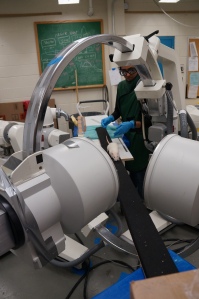
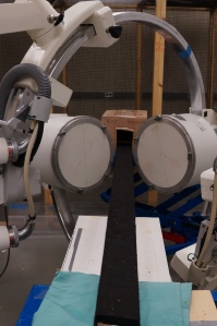
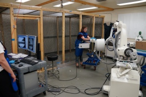
Our setup was straightforward — at the C-arms XROMM lab at Brown University, the rats walked along a plank of wood to a darkened hide box. While traversing the plank, they made their X-ray cameos in two fluoroscopes connected to hi-speed cameras filming at 250 frames per second (your iPhone camera films at 30 frames per second in normal mode). When we were finished collecting our data, the rats were CT-scanned so that we could have exact three-dimensional models of their limb bones. The most painstaking part was the several months it took to digitize each of our good trials. That is, using animation software, we had to match the bone models up to their X-ray shadows in the two calibrated fluoroscope movies. Once this was accomplished, our task turned to watching how the bones moved in three-dimensional space as well as analyzing the joint angle data that was generated.

Our basic setup for the XROMM study -- rats were trained to walk across a plank towards a dark hide box, leading them between the two videofluoroscopes.

Our basic setup for the XROMM study — rats were trained to walk across a plank towards a dark hide box, leading them between the two videofluoroscopes.

What we found both confirms previous work on small mammal locomotion, but added some interesting new insights as well. As a general rule, small mammals have a crouched posture where the elbows and knees are bent. This type of posture may aid small mammals in maneuvering around objects and keeping a lower center of gravity, which would enhance stability, especially on branches and other narrow perches. Not surprisingly and given previous work on rat locomotion, we see that these mammals do indeed walk on crouched limbs — the elbow angle, for example, never exceeded 123 degrees in full extension. By way of comparison, your elbow can be extended to 180 degrees.

A figure from my book, The Bare Bones. Note how the rat has a more crouched posture whereas the cat is more upright.

A figure from my book, The Bare Bones. Note how the rat has a more crouched posture whereas the cat is more upright.

However, we often get the impression that mammal locomotion is similar at different scales. From cats and dogs on up, it appears that the forelimbs and hindlimbs function very much as glorified pendulums. In essence, eutherian mammal locomotion is understood as mostly two-dimensional. Although rats are small and have a crouched posture, their limb bones would be presumed to follow the pendulum model.

But what the bones were doing in three-dimensions was fascinating. Both the humerus (upper arm bone) and radius (the forearm bone that aligns with your thumb) showed they were capable of long-axis rotation. Long-axis rotation is similar to the way a lathe or axle spins. Our rats’ bones certainly weren’t spinning on their long-axis, but they did show a non-trivial range of movement. A step cycle consists of a stance phase (when the hand is on the ground and forelimb is supporting the body) and a swing phase (when the hand is off the ground and the forelimb is swinging back to support the body for the next step). We found that during stance, the humerus both moves toward the midline (adducts) and rotates on its long axis towards the body. These combined movements appear to ensure that the elbow points backwards so that the forearm maintains an upright posture. During swing, the humerus moves away from the body midline (abducts) and rotates on its long axis away from the body. These combined movements seem to allow the forelimb to clear the rat’s body as the limb is brought forward to start a new step.

Lateral, ventral, and radioulnar joint views of the humerus (sea green), radius (black), and ulna (red) in a typical step cycle in Rattus norvegicus. Long-axis rotation (LAR) of the radius about the ulna (radius pronation) is shown in cranial view from the perspective of the ulna (the ulna appears to be stationary in the radioulnar joint view relative to the humerus and radius). Note radius (black) LAR relative to the ulna (red). Percentages = portion of the step cycle. Black bar in ventral view = body midline based on sternum.

Lateral (side), ventral (belly), and radioulnar joint (at the elbow) views of the humerus (sea green), radius (black), and ulna (red) in a typical step cycle in Rattus norvegicus. Long-axis rotation (LAR) of the radius about the ulna (radius pronation) is shown in cranial view (the rat is walking toward you) from the perspective of the ulna (the ulna appears to be stationary in the radioulnar joint view relative to the humerus and radius). Note radius (black) LAR relative to the ulna (red). Percentages = portion of the step cycle. Black bar in ventral view = body midline based on sternum.

MOVIE 1 – All the rats walking betwixt the fluoroscopes with their CT-scanned bones registered to the frames.

MOVIE 2 – One of our rats, “Floyd,” demonstrating a typical step cycle.

What was particularly exciting to me was that we saw, for the first time in rats, the radius pivot about the ulna! In humans, we take these movements for granted: our radius pivots around our ulna with ease, directing our palms either downward (pronated) when its shaft cross over the ulna, or upward (supinated) when its shaft rotates into parallel with the ulna. Up until now, it has been unclear if the radius could move in this way to flip the hand palm-side down in rats, or whether their hand posture was maintained via positioning of the limb in general. We now know that, indeed, the radius does move and does appear to be correlated with hand placement in rats. These movements are much more subtle than in you and I (in our rats a range of 10-30 degrees of rotation), but they appear to be correlated with pronation of the hand.

MOVIE 3 – One of our rats, “Floyd,” showing how the radius pivots on the ulna during a step cycle.

Our research has two messages. The first message is that given the similarities in the forelimb skeletons of the earliest known eutherian mammals (Juramaia and Eomaia) to those of rats, it is likely that a similar range of movements were possible in these distant relatives on our family tree. Paleontologists studying these fossils, such as Zhe-Xi Luo and colleagues (Luo et al., 2003, 2011), have already suggested these early eutherian mammals were scansorial, and our data bolster their hypothesis. These sorts of insights are helpful in constraining when particular locomotor behaviors and movements became possible and how that might have effected mammalian evolution.

The second message is that small mammal locomotion is probably not as similar to those of larger mammals as we often think, a sentiment echoed by the late Farish Jenkins (e.g., Jenkins, 1971) and by Martin Fischer and his colleagues (Fischer et al. 2002; Fisher and Blickman, 2006). Moreover, our rat data show that, at least for the forelimb, long-axis rotation plays a role in normal overground movement.

We hope our study provides another perspective on small mammal locomotion and encourages new and fruitful research in our furry friends past and present.

————————

I am grateful to my colleagues and former students for their help and work on this project. I want especially to thank Elizabeth Brainerd (Brown University). She has been a source of encouragement and a patient teacher to an old dinosaur learning new tricks, and her help with learning XROMM and on designing the experiment which led to this paper (my first foray into animal kinematics) was invaluable.

The authors of the paper (* indicates a Stockton University undergraduate)

Matthew F. Bonnan (Stockton University, Biology)
Jason Shulman (Stockton University, Physics)
*Radha Varadharajan (Stockton University, Biology)
*Corey Gilbert (Stockton University, Physics)
Mary Wilkes (Stockton University, Biology)
Angela Horner (California State University, San Berardino)
Elizabeth Brainerd (Brown University)

———————–

References

Fischer, M. S., and R. Blickhan. 2006. The tri-segmented limbs of therian mammals: kinematics, dynamics, and self-stabilization—a review. Journal of Experimental Zoology Part A: Comparative Experimental Biology 305A:935–952.

Fischer, M. S., N. Schilling, M. Schmidt, D. Haarhaus, and H. Witte. 2002. Basic limb kinematics of small therian mammals. The Journal of Experimental Biology 205:1315–38.

Jenkins, F. A. 1971. Limb posture and locomotion in the Virginia opossum (Didelphis marsupalis) and in other non-cursorial mammals. Journal of Zoology, London 165:303–315.

Luo, Z.-X., Q. Ji, J. R. Wible, and C.-X. Yuan. 2003. An Early Cretaceous Tribosphenic Mammal and Metatherian Evolution. Science 302:1934–1940.

Luo, Z.-X., C.-X. Yuan, Q.-J. Meng, and Q. Ji. 2011. A Jurassic eutherian mammal and divergence of marsupials and placentals. Nature 476:442–445.

It is official: the BFF Lab has a CT scanner

It was an exciting day for the BFF Lab — we installed and operated our Animage FIDEX CT scanner for the first time!  We selected a preserved specimen of a mudpuppy salamander (Necturus) to anoint our system.  You can see the animation below of the anterior half of the salamander’s skeleton.

Why are there large gaps between the bones of the arms, you ask?  Salamanders like the mudpuppy have thick, cartilaginous joints.  Cartilage does not typically show up in X-rays, and hence the “gaps.”

The XROMM lab at Richard Stockton University is coming together piece by piece.

We are forever grateful to lab director Justine Ciraolo and our NAMS shop director William Harron for so much help in obtaining and coordinating our receipt of this equipment.  We also want to give a big shout out to Stephen Della Ratta of Animage for his help, enthusiasm, and expertise in setting up our CT scanner.  Our training on the FIDEX scanner was thorough and friendly, and all our questions were answered.  Thank you, Stephen.

Stay tuned for more exciting news in the near future …

XROMM is coming to Stockton and the BFF Lab!

This has been working its way through the pipeline for quite awhile, but I can finally, confidently announce that the Richard Stockton College of New Jersey will house the first XROMM lab specifically focused on undergraduate research and teaching!

XROMM (X-ray Reconstruction of Moving Morphology) is a state-of-the-art technique, developed at Brown University, for visualizing rapid skeletal movement in vivo in three-dimensions.  Find out more here and here.

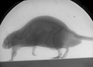
Harry, one of the rats in our trials, walking through the X-ray beams.

Harry, one of the rats in our trials, walking through the X-ray beams.

This tremendously exciting development resulted as part of a large state grant and is part of Stockton’s growing science infrastructure.  We have a second new science building on the way that will house a beautiful new vivarium and will have a custom-built XROMM lab.

The equipment we will be receiving will include hi-speed videofluoroscopes (layman’s terms: super science cool X-ray movie cameras) and a veterinary CT-scanner.

To say this is a dream come true is probably an understatement!  What it means is that we will soon have the ability to reconstruct three-dimensional moving skeletons of vertebrates for research that directly involves undergraduates.  Stay tuned to this blog and the BFF lab, and we’ll keep you posted on this exciting new development for our students and college.  My co-conspirator (eh, collaborator) Jason Shulman and I are ecstatic.

In the meantime, there are many, many people to thank.  First, Beth Brainerd, Stephen Gatesy, and the other XROMM gurus at Brown University granted me the opportunity to learn this technique through their NSF-sponsored short course.  Among the many people who have helped me understand and develop my familiarity with XROMM are David Baier and Ariel Camp, who have answered a myriad of questions.  Beth Brainerd was instrumental in this process from helping me capture my first data for analysis with Stockton undergraduate Radha Varadharajan to her generous time and assistance in understanding the specs of such a lab.  Thank you, Beth!  Angela Horner (now at California State University San Bernardino) was also instrumental in collecting our initial rat data at Brown and helping us understand how rats “tick.”

For both Jason and I, we are grateful for the on-going support and encouragement of our peers and staff at Stockton.  During the past two years, lab director Justine Ciraolo and safety officer Bob Chitren have been incredibly helpful and encouraging, and it would have been impossible to get this done without their help.  Jason and I are grateful for the support of the school of Natural Sciences and Mathematics (NAMS), to Dean Weiss, to Provost Kesselman, and President Saatkamp for supporting cutting-edge science at our college.  We are also thankful for the support and encouragement we have received from our programs, Biology and Physics, and from the generous support of the Provost and Grants Office for internal grants that have placed us in this exciting position.

We must make a special mention of John Rokita and the animal lab staff for keeping our animals happy and healthy, and the Institutional Animal Care and Usage Committee (IACUC) here at Stockton for overseeing our animal research.  Again, the NAMS laboratory staff are to be thanked for all of their continuing help in making such exciting STEM experiences possible for our students.

Finally, Jason and I are delighted that we can bring this caliber of research to our students at Stockton.  It will allow us to expand on our locomotion research using optical tracking, and give students pursuing a wide range of careers in the sciences a rare opportunity to learn about the living skeleton in action.  Most importantly, the XROMM lab will expand Stockton’s already strong history of producing New Jersey STEM majors.

We will blog and tweet about the progress of the XROMM lab setup and keep you informed about how it is all coming together over the next several months.  Stay tuned!

The Visit to Brown University

Radha Varadharajan at Brown with Beth Brainerd.

Radha Varadharajan at Brown with Beth Brainerd.

After spending five valuable days at Brown University, Dr. Matthew Bonnan and I learned a great deal about C-arm fluoroscopes and with XROMM technology. The early stages involved getting accustomed to the protocols of working with the fluoroscopes. This step was pivotal for the machines emit an innocuous but not neglible amount of radiation to capture the motions of our rats: Pink, Floyd, Evan, Rudy, Harry, and Taylor.  Personally, the pinnacle of the visit to Brown can be identified as the days the rats walked across the beam. With much guidance from both me and Dr. Bonnan, our furry test subjects were cajoled across the plank or dowel. Although Dr. Bonnan was the primary coaxer of our scampering participants, I was also able to give a hand in guiding them.

Within a few days into the visit, I was amazed with the advanced technologies at Brown. Comprehending the process of how the fluoroscopes operated was especially intriguing. Because I was able to accompany Dr. Bonnan on this trip to Brown University, not only did I understand the innovational technology that is XROMM, but I was also able to contribute in the smallest way possible in understanding the evolution of forelimb posture.

Here is Radha coaxing Pink the Rat through the X-ray beams.

Here is Radha coaxing Pink the Rat through the X-ray beams.

I would like to conclude this post by expressing my utmost gratitude to numerous individuals that allowed for my collaboration. My involvement in this educational visit would not have been feasible without the generous contribution of Dr. Robert Fine; Dr. Fine’s munificent gesture solely funded my trip. I would also like to thank Dr. Elizabeth Brainerd and Dr. Angela Horner for their guidance. Finally, I would like acknowledge Dr. Bonnan for his unceasing support.  Without his patience, I would not have been able to discover the numerous benefits of researching in such a compelling field.

Rodents of usual size and their moving skeletons

Harry, one of the rats in our trials, walking through the X-ray beams.

Harry, one of the rats in our trials, walking through the X-ray beams.

The past week at Brown University’s C-arms XROMM lab was so busy I haven’t had a moment to post about our research experiences until now.  If you’re just catching up, please see my previous post on our setup.

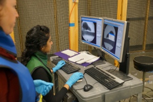
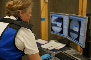
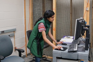
This was certainly a new but fascinating experience both for me and my student, Radha.  With help from Dr. Beth Brainerd and Dr. Angela Horner, we learned how to coax the rats to walk a plank of wood between the two X-ray emitting “cans” of the positioned C-arm fluoroscopes.  At one end of the room is a bank of two computers connected to each high-speed camera and C-arm.  When the rats were doing what we were interested in, a push of a floor pedal turned on the X-rays and recorded the ensuing stream of images which were then converted into standard computer movies.

Walk the plank - each rat walked across this plan between the C-arm fluoroscopes to a hidey-hole box we nick-named the Rat Haven.

Walk the plank – each rat walked across this plank between the C-arm fluoroscopes to a hidey-hole box we nick-named the Rat Haven.

Dr. Brainerd helping Radha and I to capture the X-ray data.

Dr. Brainerd helping Radha and I to capture the X-ray data.

Radha Varadharajan at C-arms lab

Here is Radha Varadharajan capturing and recording the X-ray movies that will be the foundation of our study.

Angela Horner has been working with rats for years, and her experience in motivating these little mammals was a godsend — from Wednesday to Thursday, Radha and I learned from her experience and were able to collect loads of data that will allow us to begin reconstructing their locomotor and postural movements in 3-D.

Here, Dr. Angela Horner is motivating the rat Harry to walk the plank through the X-ray beams.

Here, Dr. Angela Horner is motivating the rat Harry to walk the plank through the X-ray beams.

Radha and I both had opportunities to coax the rats across the plank to the Rat Haven as well.  You will notice we named our rats.  Two of them were dubbed Pink and Floyd as a nod to one of my favorite bands who also featured cartoon rats in their backdrop movie for “Welcome to the Machine.”  Yeah, we’re geeky like that.

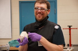
Here I am holding one of the rats we named Evan.  Evan was a bit "lazy," but ended up being great at walking a narrow dowel, helping us to see forearm movements in detail.

Here I am holding one of the rats we named Evan. Evan was a bit “lazy,” but ended up being great at walking a narrow dowel, helping us to see forearm movements in detail.

Here is Radha coaxing Pink the Rat through the X-ray beams.

Here, Radha is coaxing Pink the Rat through the X-ray beams.

Want to see a sneak-peak of the end result of our labors?  Here is one clip of Harry the Rat.

We are especially grateful for all the help we had this past week, and among many others Erika Giblin and Ariel Camp were invaluable in providing access and assistance with all of our XROMM issues.  Thank you everyone!

An old dinosaur learning new tricks

Okay, so I’m the “old dinosaur” here, although I was informed recently that I could still pass as a graduate student.

I am happy to report that I am back on the campus of Brown University this week with one of my undergraduates, Radha Varadharajan, to begin what I hope to be the first in a long series of studies on the evolution of amniote (reptile, bird, mammal) forelimb posture.  We (my “rat pack” students and I) are using the XROMM technology I have detailed here on this blog to understand how the three-dimensional movements of the forelimb bones of rats actually occur.  The long-term goal of this initial study is to document how these movements facilitate hand placement and posture, and how these details of locomotion are related to bone shape.  My ultimate goal is to use the somewhat primitive forelimb posture of rats as a template to understand how some early fossil mammals may have moved.

Today, Radha and I, under the tutelage of Dr. Elizabeth Brainerd, began the process of setting up the so-called C-arm fluoroscopes that will allow us to take calibrated X-ray movies of a number of rats as they walk, run, and perhaps do other activities that we happen to capture.  This was especially exciting and informative for me, because these are the “new tricks” this “old dinosaur” wants to learn.  Tomorrow, we begin in earnest filming the skeletal movements of the rats.

You will notice in the pictures posted here that Radha and I are suited up in lead aprons and thyroid collars because, as you might anticipate, we do not want to expose ourselves to X-ray radiation during the data capture.  In fact, she and I have participated in numerous safety trainings and tests to ensure we stay safe.

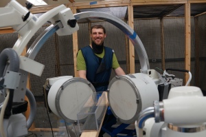
Here I am behind the two C-arm fluoroscopes.  In front of the scopes, you can spy the wooden plank walk-way for the rats, and an acrylic box that the rats will walk or run through in the vicinity of the X-ray fields.

Here I am behind the two C-arm fluoroscopes. In front of the scopes, you can spy the wooden plank walk-way for the rats, and an acrylic box that the rats will walk or run through in the vicinity of the X-ray fields.

Here is Radha learning x-ray capture at the Brown C-arms lab.

Here is Radha learning x-ray capture at the Brown C-arms lab.

We also spent time today with Dr. David Baier learning how to set up what is called a rig in the MAYA software program that will later animate the skeletons of the rats we film.  Essentially, a rig in this case means creating a joint system that can be calibrated with the X-ray films and “attached” to the 3-D bone geometry from CT-scans of the rats used in the study.  I further shook some of the rust out of my head reviewing and practicing how to import calibrated data from X-ray digital movies and syncing them with 3-D bone geometry — skills I first acquired almost one year ago during Brown’s 2012 XROMM course.

All of this setup and learning is key for me and my students, not only because we want to do the science right, but also for other reasons I shall divulge in future posts.

Everyone at Brown has once again been incredibly helpful, and I am especially indebted to Dr. Brainerd for her encouragement and help over the past year with XROMM.

Please stay tuned … this week promises to get more interesting …

The “Rat Pack” Succeeds

A lot has happened in the Bonnan Lab at Stockton these past few months.

First, the “Rat Pack” as I fondly call them (Evan Drake, Kadeisha Pinkney, and Radha Varadharajan), presented their research proposals for 3-D rat locomotion and kinematics to the Northeastern Regional Vertebrate Evolution Symposium (NERVES) on March 22nd, 2013, at the New York Institute of Technology (NYIT) College of Osteopathic Medicine.  Their talks were very well received and we had excellent suggestions from colleagues and scholars.  I was especially proud of these undergraduates because they were able to give technical talks to a scientific audience having only worked with me for a few months on their projects. Bravo!

A special thank you to the Symposium’s organizers, Drs. Brian Beatty and Matthew Mihlbachler!

You can read their NERVES talk titles below.

Next, they put their collective heads together and, with my input, created a very nice poster for the 2013 NAMS Research Symposium at Stockton.

You can read their NAMS research abstract below as well.

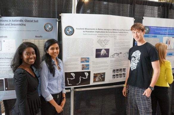
The "Rat Pack" presented their preliminary research on rat locomotion to the 2013 NAMS Research Symposium.  Left to right: Kadeisha Pinkney, Radha Varajharadan, and Evan Drake.

The “Rat Pack” presented their preliminary research on rat locomotion to the 2013 NAMS Research Symposium. Left to right: Kadeisha Pinkney, Radha Varadharajan, and Evan Drake.

Kadeisha (left) and Radha (right) explain the joys of rat locomotion to interested students.

The “Rat Pack” attracted quite a crowd.

Last, but not least, Radha Varadharajan received the first Robert L. Fines scholarship awarded at Stockton, April 26, 2013, for her work on this research and her future career goals in veterinary medicine.  Dr. Fines is a former Stockton alumnus (1975) and is one of the premiere M.D. researchers successfully fighting pancreatic cancer.  He is the Herbert Irving Associate Professor of Medicine in the Division of Medical Oncology at the Columbia University College of Physicians & Surgeons in New York, New York.  I am honored and proud that Radha has received this award from such a prestigious alumnus.

The scholarship will allow Radha to travel with me to Brown University the week of May 20-24, where we will work with Dr. Elizabeth Brainerd and colleagues X-ray filming the rats walking, running, and landing at their XROMM C-arms facility.  Stay tuned and we’ll keep you posted on our time and research activities while at Brown.

Finally, I must acknowledge the help of our campus veterinarians, Drs. Ralph Werner and Mary Wilkes, for their efforts in helping me with the rats, as well as our animal caretaker, John Rokita, for his constant help and suggestions on rodent protocols and biology.

I feel truly grateful to have made such a jump to a new college and to already be surrounded by supportive faculty, eager students, and the chance to pursue 3-D kinematics research.

NERVES Talk Titles

Varadharajan, Radha and Bonnan, Matthew F. 2013. Exploring 3-D long bone kinematics in the White Rat (Rattus norvegicus) as a model for inferring forelimb posture in early mammals: Contribution of the scapula.>

Pinkney, Kadeisha and Bonnan, Matthew F. 2013. Exploring 3-D long bone kinematics in the White Rat (Rattus norvegicus) as a model for inferring forelimb posture in early mammals: Contribution of the humerus.

Drake, Evan and Bonnan, Matthew F. 2013. Exploring 3-D long bone kinematics in the White Rat (Rattus norvegicus) as a model for inferring forelimb posture in early mammals: Contribution of the radius and ulna.

NAMS RESEARCH SYMPOSIUM ABSTRACT

Forelimb movements in Rattus norvegicus (white rat) and their relationship to pronation: implications for early mammal forelimb posture

Varadharajan, Radha; Pinkney, Kadeisha; Drake, Evan; and Bonnan, Matthew F.


Rattus norvegicus (the white rat) is a therian mammal with a forelimb morphology similar to that of early non-cursorial mammals. Currently, early mammal limb posture is controversial, with reconstructions ranging from sprawling to parasagittal. With this current ambiguity, the study of forelimb shape and movements in R. norvegicus may provide a model to infer the locomotor patterns of earlier mammals.  Previous research, most notably by Jenkins (1971, 1974), indicates that the forelimb posture of rats does not follow simple, pendulum-like mechanics but rather a more complex, less-upright range of movement. For the first time, we will study the 3-D morphology and kinematics of the forelimb in R. norvegicus by utilizing three-dimensional moving X-ray animations generated through the XROMM (X-ray Reconstruction of Moving Morphology) technique.  Specifically, we will focus on the three-dimensional movements of the scapula, humerus, and antebrachium (radius and ulna), and their combined contribution to pronation (placing the hand palm-side down).  To this end, we will test three interdependent hypotheses on the contribution of each of these limb segments to pronation. For the scapula, we examined the serratus anterior, supraspinatous, infraspinatous, spinotrapezius, acromiotrapezius, and rhomboids major and minor. Data gathered on the rat scapula through literature and dissection lead to the hypothesis that this element contributes in a significant way to pronation. Specific features of the humerus distinguish the parasagittal from the sprawling stance in early fossil mammals: degree of torsion, condylar structures of the elbow joint, width of the intertubercular groove, and the relative sizes of the lesser and greater tubercles.  These features are associated with major locomotor muscles such as the pectoralis major and minor, deltoids, and pronator teres. We hypothesize that the humerus will contribute in a significant way to the pronation of the hand in the white rat. In humans, the radius can rotate about the ulna to pronate the hand because these elements are bowed, creating the space necessary to allow such movements. The pronator teres and pronator quadratus pull on the radius and rotate it about the ulna, whereas the supinator and biceps brachii act as antagonists to return the bones to a parallel position. Unlike humans, the radius and ulna of Rattus norvegicus fit tightly together like two spoons stacked together, with little, if any, space available in which the radius can rotate about the ulna.  Moreover, the pronator quadratus has not yet been described or identified in our rat dissections. Instead, the radius and ulna appear “fused” by the interosseous membrane and rendered incapable of supination. Rats have little need to supinate the forelimb because the forelimbs are primarily used in locomotion. It is therefore hypothesized that all pronation and supination occur in the humerus and scapula in rats because the radius and ulna are in such close proximity to each other that we infer they participate little, if any, to pronation.  After we capture the three-dimensional movements of these bones with XROMM, we will test our hypotheses and perhaps gain insight into the posture of early mammals.

XROMM Days 3-5: Data, data, data

Again, if you’re just tuning in to this thread on XROMM, please refer to my previous posts on what XROMM is, why I am excited about using it, and what I have done so far, including 3-D animating a chewing pig.

The past few days have seen us busy and social.  We’ve now learned to do scientific rotoscoping, or the art of properly aligning three-dimensional, anatomically-accurate models of vertebrates with two X-ray movies shot from different angles.  I have previously described the basic steps involved in making these XROMM movies and coordinating and calibrating the X-ray movies, so I refer you to my previous post on this.

Unlike the method of XROMM I described previously that uses bone markers to synch the moving images and the bones, in scientific rotoscoping you manually align bones frame by frame with x-ray movies.  It can be tedious at times, but essentially it is, to quote Steve Gatesy, quoting Ken Dial, fitting a digital key (the bones) into a visual lock (the X-ray movies).  In other words, you are posing the skeleton in three-dimensional coordinates based on the constraints imposed by X-ray movies.  Put simply, if your digital skeleton deviates from the reality of the X-ray movies, you are doing something wrong.

What is perhaps most important to emphasize in all of this is that because you are fitting bones into a virtual reality space of what happened when the animal was filmed, you can now recover data about skeletal and joint movements.  That’s right: you can actually retrieve reliable, repeatably scientific data on gait and movement from these 3-D animations.  For example, you assign joint markers and then measure how much the mouth of a pig opens, or how much rotation is happening in a bird knee, or even how parts of a fish skull move in relation to the skeleton and muscles.

So, you are not just making a nice skeleton movie — you are recovering what would normally be unrecoverable data.  The sort of data that allows you to more objectively describe what happens in living vertebrates, and hopefully, data that can be used as a baseline constraint for limiting what fossil vertebrates may or may not have been capable of.

All in all, this has been a great experience.  I wish to thank Beth Brainerd, Steve Gatesy, David Baier, Ariel Camp, Sabine Moritz, and Erika Giblin for all their help, information, and assistance this week.  It has been a pleasure.

XROMM Day 2: This little piggy bites … in 3-D

Again, if you missed previous posts on XROMM, please read those first for better context of the discussion that follows: see what XROMM is all about, why I’m excited to be learning it, and what I’ve already done.

On our second day in the course, we took the next step to synch two different, simultaneous X-ray movies of a mini-pig eating with a three-dimensional model of its skull in the MAYA program.  These X-ray films were taken on the C-arm x-ray machines I mentioned in my previous post by Dr. Beth Brainerd and colleagues, and we were essentially learning by replicating their process.

Dr. B. and the Mini-Pig

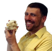
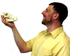
Me and mini-pig’s skull. The original movie was filmed back in 2006, so the mini-pig that stars in the X-rays has since passed on to piggy heaven. Here I am holding the actual skull of the animal that you will see in the animation.

In XROMM, you essentially have a work-flow like this:

  1. Film an animal behavior from two directions using calibrated X-ray cameras.  The animal usually has tiny, spherical beads implanted surgically into a few of the bones of interest prior to the filming.  As an important note here, all such films and surgeries are done under strict animal welfare protocols and the animals are not harmed: the X-ray dosages are as low or lower than that of humans exposed to x-rays for diagnosis, and the beads are tiny and biologically inert.
  2. After filming the animal’s behavior, the skeleton’s three-dimensional geometry is often CT-scanned from the animal.
  3. The CT-scan bone data are converted into geometrically-accurate 3-D representations of the bones of interest.
  4. In MAYA and MATLAB, the films from the two x-ray cameras are virtually “projected,” and the beads implanted into the animal subject show up as little dots.  These dots show up as little spheres in the CT-scanned bones you import into the MAYA program.
  5. Things then get more technical, but suffice it to say that the beads you see in the X-ray films and the spheres in the CT bones are synched.  The movements of the beads (spheres) are digitized in three-dimensions calibrated from the two camera views, and then the virtual bones are “cemented” to these spheres.  Then, as the spheres move, the bones follow, and what you get is a three-dimensional reconstruction of the 2-D x-ray films!

Here is a screen shot of the animation made in XROMM today.

Mini-Pig X-ray and Reconstruction

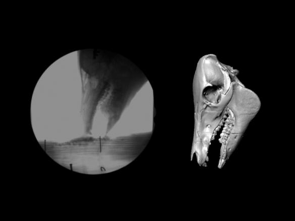
What you get from XROMM: to the left is one of the two X-ray films, and to the right is the 3-D skull reconstruction of the mini-pig.

Here is an animation of what you get — I still can’t believe we can do this!

Dr. B. Contemplates the Mini-Pig

Dr. B. Contemplates the mini-pig skull.

And we’re not even done yet.  Stay tuned …

XROMM Day 1: Pig heads and C-arms

If you’re just tuning in, you may want to read my previous posts on what XROMM is and why I am thrilled to be learning this technique.

Today was a good, busy, productive day at the XROMM course.  Never let it be said that “simply” synching X-ray “movies” (cineradiography) is easy!  The students in the course are learning how to animate the three-dimensional jaw movements of a mini-pig, based on the research of Dr. Beth Brainerd and colleaguesThe XROMM site provides movies and animations of what we are attempting to duplicate.

You don’t simply take an X-ray movie and then transpose that into a 3-D animation, of course.  If you’re going to match up the three-dimensional models of the mini-pig skull to the chewing motions recorded on as cineradiographs, you have to get the animation program (MAYA) to synch with the frames of these movies.  And that involves a number of techniques including correcting for distortion (the tube that transmits the X-ray images to the camera has convex ends, which give the original footage a fish-eye lens distortion) and “registering” the simultaneous side and top or bottom views of the moving animal to virtual cameras and screens in MAYA.

In science, the tedious parts come from making absolutely sure you are doing everything to account for error and noise in your data.  This usually pays off with dividends in the end, but getting there is the hard work. Its not enough in this case to do all the technical things necessary simply to capture a moving animal’s skeleton in two planes.  Then you have to spend hours and days matching that raw data to your virtual skeleton — and all for a sequence of maybe 2-20 seconds in length.

Today we also were able to visit the two XROMM facilities at Brown.

Dr. Bonnan at XROMM facility

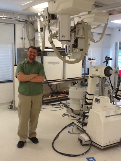
Dr. Bonnan in one of the two labs of the XROMM facility a Brown University. Note the two large, mobile X-ray machines to Dr. Bonnan’s left.

Physics came back to haunt me today, as part of doing the science of XROMM correctly and safely is putting knowledge of photons and radiomagnetic waves to good use. Physics was always a tough subject for me, but it is amazing what you can learn and apply when you really want to do something and when doing it incorrectly will result in long-term injuries from X-ray irradiation!

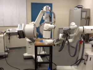
One of the rooms had mobile C-arm X-ray machines that demonstrate very well the basic concept of what you do when you capture the motions of an animal.

Small C-arms at XROMM

Two small C-arm X-ray machines in one of the XROMM labs, positioned perpendicular to one another. One is facing left, and the other is pointed away from you in this photo.

All of the XROMM faculty and staff have been wonderful and saintly in their patience with us as we learn a technique that is often as frustrating to learn as it is to explain many times over and over again.  A big thank you for their patience and help today … and I think my eyes are starting to uncross now.

To give you an idea of the time investment necessary to convert 2-D cineradiographs into 3-D moving models, consider this: we spent most of our time today simply registering points and synching virtual camera views … there has been no animation yet!

I see great potential for research and student involvement with XROMM, and I’m looking forward to having a chewing mini-pig skull in the next few days.

Stay tuned …